 Movie
Movie Controller

[English] 日本語
 Yorodumi
- EMDB-14021: Cryo-ET of Magnetospirillum gryphiswaldense delta-popZ overproduc... -
+ Open data

- Basic information
| Entry | Database: EMDB / ID: EMD-14021 | |||||||||
|---|---|---|---|---|---|---|---|---|---|---|
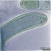
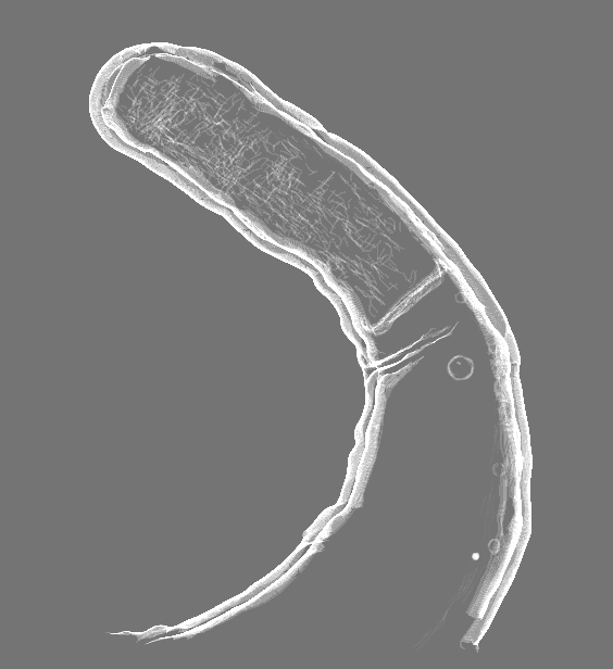
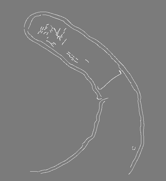
| Title | Cryo-ET of Magnetospirillum gryphiswaldense delta-popZ overproducing PopZ-Mgr | |||||||||
 Map data Map data | Tomogram of Magnetospirillum gryphiswaldense delta-popZ overproducing PopZ-Mgr | |||||||||
 Sample Sample |
| |||||||||
 Keywords Keywords | PopZ / Alphaproteobacteria / cytoskeleton / polarity / CELL CYCLE | |||||||||
| Biological species |  Magnetospirillum gryphiswaldense MSR-1 (magnetotactic) Magnetospirillum gryphiswaldense MSR-1 (magnetotactic) | |||||||||
| Method | electron tomography / cryo EM | |||||||||
 Authors Authors | Toro-Nahuelpan M / Plitzko JM / Schueler D / Pfeiffer D | |||||||||
| Funding support |  Germany, 2 items Germany, 2 items
| |||||||||
 Citation Citation | Journal: J Mol Biol / Year: 2022 Title: In vivo Architecture of the Polar Organizing Protein Z (PopZ) Meshwork in the Alphaproteobacteria Magnetospirillum gryphiswaldense and Caulobacter crescentus. Authors: Mauricio Toro-Nahuelpan / Jürgen M Plitzko / Dirk Schüler / Daniel Pfeiffer / Abstract: The polar organizing protein Z (PopZ) forms a polar microdomain that is inaccessible to larger macromolecules such as ribosomes, and selectively sequesters proteins crucial for cell cycle control and ...The polar organizing protein Z (PopZ) forms a polar microdomain that is inaccessible to larger macromolecules such as ribosomes, and selectively sequesters proteins crucial for cell cycle control and polar morphogenesis in various Alphaproteobacteria. However, the in vivo architecture of this microdomain has remained elusive. Here, we analyzed the three-dimensional ultrastructural organization of the PopZ network in Magnetospirillum gryphiswaldense and Caulobacter crescentus by Volta phase plate cryo-electron tomography, which provides high spatial resolution and improved image contrast. Our results suggest that PopZ forms a porous network of disordered short, flexible, and branching filaments. | |||||||||
| History |
|
- Structure visualization
| Movie |
 Movie viewer Movie viewer |
|---|---|
| Supplemental images |

- Downloads & links
-EMDB archive
| Map data | emd_14021.map.gz | 34.2 MB | EMDB map data format | |
|---|---|---|---|---|
| Header (meta data) | emd-14021-v30.xmlemd-14021.xml | 12.9 KB 12.9 KB | Display Display | EMDB header |
| Images |  emd_14021.png emd_14021.png | 39.7 KB | ||
| Masks | emd_14021_msk_1.map | 40.1 MB | Mask map | |
| Filedesc metadata | emd-14021.cif.gz | 4.8 KB | ||
| Archive directory |  http://ftp.pdbj.org/pub/emdb/structures/EMD-14021ftp://ftp.pdbj.org/pub/emdb/structures/EMD-14021 http://ftp.pdbj.org/pub/emdb/structures/EMD-14021ftp://ftp.pdbj.org/pub/emdb/structures/EMD-14021 | HTTPS FTP |
-Validation report
| Summary document | emd_14021_validation.pdf.gz | 330.2 KB | Display | EMDB validaton report |
|---|---|---|---|---|
| Full document | emd_14021_full_validation.pdf.gz | 329.7 KB | Display | |
| Data in XML | emd_14021_validation.xml.gz | 5.1 KB | Display | |
| Data in CIF | emd_14021_validation.cif.gz | 6.1 KB | Display | |
| Arichive directory | https://ftp.pdbj.org/pub/emdb/validation_reports/EMD-14021ftp://ftp.pdbj.org/pub/emdb/validation_reports/EMD-14021 | HTTPS FTP |
-Related structure data
| Related structure data | C: citing same article ( |
|---|---|
| EM raw data | EMPIAR-10885 (Title: In vivo architecture of the polar organizing protein Z (PopZ) meshwork in the Alphaproteobacteria Magnetospirillum gryphiswaldense and Caulobacter crescentus Data size: 6.3 Data #1: Volta phase plate cryo-electron tomography of Magnetospirillum gryphiswaldense (wild-type) overproducing PopZ-Mgr [reconstructed volumes] Data #2: Volta phase plate cryo-electron tomography of Magnetospirillum gryphiswaldense delta-mamK overproducing PopZ-Mgr [reconstructed volumes] Data #3: Volta phase plate cryo-electron tomography of purified PopZ-Mgr [reconstructed volumes] Data #4: Cryo-electron tomography of Magnetospirillum gryphiswaldense delta-popZ overproducing PopZ-Mgr [reconstructed volumes] Data #5: Volta phase plate cryo-electron tomography of Caulobacter crescentus NA1000 [reconstructed volumes] Data #6: Volta phase plate cryo-electron tomography of Caulobacter crescentus NA1000 overproducing PopZ-Cc [reconstructed volumes] Data #7: Cryo-electron tomography of Magnetospirillum gryphiswaldense delta-popZ [reconstructed volumes] Data #8: Volta phase plate cryo-electron tomography of Magnetospirillum gryphiswaldense delta-mamK-mamY [reconstructed volumes]) |
-Links
| EMDB pages | EMDB (EBI/PDBe) / EMDataResource |
|---|
-Map
| File | Download / File: emd_14021.map.gz / Format: CCP4 / Size: 40.1 MB / Type: IMAGE STORED AS SIGNED BYTE | ||||||||||||||||||||||||||||||||||||||||||||||||||||||||||||||||||||
|---|---|---|---|---|---|---|---|---|---|---|---|---|---|---|---|---|---|---|---|---|---|---|---|---|---|---|---|---|---|---|---|---|---|---|---|---|---|---|---|---|---|---|---|---|---|---|---|---|---|---|---|---|---|---|---|---|---|---|---|---|---|---|---|---|---|---|---|---|---|
| Annotation | Tomogram of Magnetospirillum gryphiswaldense delta-popZ overproducing PopZ-Mgr | ||||||||||||||||||||||||||||||||||||||||||||||||||||||||||||||||||||
| Voxel size | X=Y=Z: 31.32 Å | ||||||||||||||||||||||||||||||||||||||||||||||||||||||||||||||||||||
| Density |
| ||||||||||||||||||||||||||||||||||||||||||||||||||||||||||||||||||||
| Symmetry | Space group: 1 | ||||||||||||||||||||||||||||||||||||||||||||||||||||||||||||||||||||
| Details | EMDB XML:
CCP4 map header:
| ||||||||||||||||||||||||||||||||||||||||||||||||||||||||||||||||||||
-Supplemental data
-Mask #1
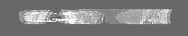
| File | emd_14021_msk_1.map | ||||||||||||
|---|---|---|---|---|---|---|---|---|---|---|---|---|---|
| Projections & Slices |
| ||||||||||||
| Density Histograms |
 Z
Z Y
Y X
X


- Sample components
-Entire : Magnetospirillum gryphiswaldense delta-popZ overproducing PopZ-Mgr
| Entire | Name: Magnetospirillum gryphiswaldense delta-popZ overproducing PopZ-Mgr |
|---|---|
| Components |
|
-Supramolecule #1: Magnetospirillum gryphiswaldense delta-popZ overproducing PopZ-Mgr
| Supramolecule | Name: Magnetospirillum gryphiswaldense delta-popZ overproducing PopZ-Mgr type: cell / ID: 1 / Parent: 0 Details: Individual segmented structures are depicted in different colors in the EMDB entry image and in the corresponding publication: Putative PopZ filaments are purple. Magnetite crystals are red, ...Details: Individual segmented structures are depicted in different colors in the EMDB entry image and in the corresponding publication: Putative PopZ filaments are purple. Magnetite crystals are red, magnetosome membrane vesicles are yellow, and the cellular envelope inner and outer membranes are depicted in blue. MamK filaments are green. |
|---|---|
| Source (natural) | Organism: Magnetospirillum gryphiswaldense MSR-1 (magnetotactic) |
-Supramolecule #2: PopZ
| Supramolecule | Name: PopZ / type: organelle_or_cellular_component / ID: 2 / Parent: 1 Details: The polar organizing protein Z (PopZ) forms a polar microdomain that is inaccessible to larger macromolecules such as ribosomes, and selectively sequesters proteins crucial for cell cycle ...Details: The polar organizing protein Z (PopZ) forms a polar microdomain that is inaccessible to larger macromolecules such as ribosomes, and selectively sequesters proteins crucial for cell cycle control and polar morphogenesis in various Alphaproteobacteria. In the present Magnetospirillum gryphiswaldense delta-popZ strain (lacking the endogenous popZ gene) PopZ overproduction was achieved via genomic insertion of a Tn5-Ptet-based popZ overexpression cassette. |
|---|---|
| Source (natural) | Organism: Magnetospirillum gryphiswaldense MSR-1 (magnetotactic) |
-Supramolecule #3: Magnetosomes
| Supramolecule | Name: Magnetosomes / type: organelle_or_cellular_component / ID: 3 / Parent: 1 Details: Magnetosomes are membranous organelles containing nanometer-sized crystals of magnetite present in magnetotactic bacteria. |
|---|---|
| Source (natural) | Organism: Magnetospirillum gryphiswaldense MSR-1 (magnetotactic) |
-Supramolecule #4: magnetite crystals
| Supramolecule | Name: magnetite crystals / type: organelle_or_cellular_component / ID: 4 / Parent: 3 |
|---|---|
| Source (natural) | Organism: Magnetospirillum gryphiswaldense MSR-1 (magnetotactic) |
-Supramolecule #5: magnetosome membrane vesicles
| Supramolecule | Name: magnetosome membrane vesicles / type: organelle_or_cellular_component / ID: 5 / Parent: 3 |
|---|---|
| Source (natural) | Organism: Magnetospirillum gryphiswaldense MSR-1 (magnetotactic) |
-Supramolecule #6: MamK
| Supramolecule | Name: MamK / type: organelle_or_cellular_component / ID: 6 / Parent: 1 Details: The filament-forming bacterial actin MamK found in magnetotactic bacteria is important for organizing magnetosome organelles into chains that are used for navigation along geomagnetic field lines. |
|---|---|
| Source (natural) | Organism: Magnetospirillum gryphiswaldense MSR-1 (magnetotactic) |
-Supramolecule #7: Cellular envelope
| Supramolecule | Name: Cellular envelope / type: organelle_or_cellular_component / ID: 7 / Parent: 1 / Details: inner and outer membranes |
|---|---|
| Source (natural) | Organism: Magnetospirillum gryphiswaldense MSR-1 (magnetotactic) |
-Experimental details
-Structure determination
| Method | cryo EM |
|---|---|
 Processing Processing | electron tomography |
| Aggregation state | cell |
-Sample preparation
| Buffer | pH: 7 / Details: modified flask standard medium (FSM) |
|---|---|
| Grid | Model: Quantifoil / Material: COPPER / Support film - Material: CARBON / Support film - topology: HOLEY / Pretreatment - Type: GLOW DISCHARGE |
| Vitrification | Cryogen name: ETHANE Details: The mixture was blotted and embedded in vitreous ice by plunge freezing into liquid ethane (< - 170 C). The grids were stored in sealed boxes in liquid nitrogen until used.. |
| Sectioning | Other: NO SECTIONING |
| Fiducial marker | Manufacturer: Sigma-Aldrich / Diameter: 15 nm |
- Electron microscopy
| Microscope | FEI POLARA 300 |
|---|---|
| Image recording | Film or detector model: GATAN K2 SUMMIT (4k x 4k) / Detector mode: COUNTING / Average electron dose: 1.5 e/Å2 Details: The cumulative electron dose during the tilt series was kept below 150 e- A-2. |
| Electron beam | Acceleration voltage: 300 kV / Electron source:  FIELD EMISSION GUN FIELD EMISSION GUN |
| Electron optics | Illumination mode: FLOOD BEAM / Imaging mode: DARK FIELD / Nominal defocus max: 5.0 µm / Nominal defocus min: 5.0 µm |
| Experimental equipment |  Model: Tecnai Polara / Image courtesy: FEI Company |
-Image processing
| Final reconstruction | Number images used: 121 |
|---|
