endonucleolytic cleavage to generate mature 3'-end of SSU-rRNA from (SSU-rRNA, 5.8S rRNA, LSU-rRNA) / endonucleolytic cleavage in ITS1 to separate SSU-rRNA from 5.8S rRNA and LSU-rRNA from tricistronic rRNA transcript (SSU-rRNA, 5.8S rRNA, LSU-rRNA) / maturation of SSU-rRNA from tricistronic rRNA transcript (SSU-rRNA, 5.8S rRNA, LSU-rRNA) / small-subunit processome / rRNA processing / ribosomal small subunit assembly / ribosomal small subunit biogenesis / small ribosomal subunit rRNA binding / cytosolic small ribosomal subunit / cytosolic large ribosomal subunit ...endonucleolytic cleavage to generate mature 3'-end of SSU-rRNA from (SSU-rRNA, 5.8S rRNA, LSU-rRNA) / endonucleolytic cleavage in ITS1 to separate SSU-rRNA from 5.8S rRNA and LSU-rRNA from tricistronic rRNA transcript (SSU-rRNA, 5.8S rRNA, LSU-rRNA) / maturation of SSU-rRNA from tricistronic rRNA transcript (SSU-rRNA, 5.8S rRNA, LSU-rRNA) / small-subunit processome / rRNA processing / ribosomal small subunit assembly / ribosomal small subunit biogenesis / small ribosomal subunit rRNA binding / cytosolic small ribosomal subunit / cytosolic large ribosomal subunit / cytoplasmic translation / rRNA binding / structural constituent of ribosome / ribosome / translation / ribonucleoprotein complex / mRNA binding / nucleolus / RNA binding / metal ion binding / nucleus / cytosol Similarity search - Function
: / Ribosomal protein S26e signature. / Ribosomal protein L41 / Ribosomal protein L41 / Ribosomal protein S21e, conserved site / Ribosomal protein S21e signature. / Ribosomal protein S26e / Ribosomal protein S26e superfamily / Ribosomal protein S26e / Ribosomal protein S5, eukaryotic/archaeal ...: / Ribosomal protein S26e signature. / Ribosomal protein L41 / Ribosomal protein L41 / Ribosomal protein S21e, conserved site / Ribosomal protein S21e signature. / Ribosomal protein S26e / Ribosomal protein S26e superfamily / Ribosomal protein S26e / Ribosomal protein S5, eukaryotic/archaeal / Ribosomal protein S21e / Ribosomal protein S21e superfamily / Ribosomal protein S21e / Ribosomal protein L19, eukaryotic / Ribosomal protein S2, eukaryotic / Ribosomal protein L19/L19e conserved site / Ribosomal protein L19e signature. / Ribosomal protein S30 / Ribosomal protein S30 / Ribosomal protein S8e subdomain, eukaryotes / : / Ribosomal protein S7e signature. / Ribosomal protein S17e, conserved site / Ribosomal protein S17e signature. / Ribosomal protein S2, eukaryotic/archaeal / 60S ribosomal protein L19 / Ribosomal protein S3Ae, conserved site / Ribosomal protein S3Ae signature. / Ribosomal protein S8e, conserved site / Ribosomal protein S8e signature. / Ribosomal protein S27e signature. / Ribosomal protein S6, eukaryotic / Ribosomal protein S17e / Ribosomal protein S17e-like superfamily / Ribosomal S17 / : / : / Ribosomal protein L19e, C-terminal domain / Ribosomal_L19e / Ribosomal protein L19/L19e / Ribosomal protein L19/L19e, domain 1 / Ribosomal protein L19/L19e superfamily / Ribosomal protein L19e, N-terminal domain / 40S ribosomal protein S1/3, eukaryotes / 40S ribosomal protein S11, N-terminal / Ribosomal_S17 N-terminal / Ribosomal protein S7e / Ribosomal protein S7e / : / Ribosomal S24e conserved site / Ribosomal protein S24e signature. / Ribosomal protein S6/S6e/A/B/2, conserved site / Ribosomal protein S6e signature. / Ribosomal protein S17, archaeal/eukaryotic / Ribosomal protein S23, eukaryotic/archaeal / Ribosomal protein S8e / Ribosomal protein S27 / Ribosomal protein S27, zinc-binding domain superfamily / Ribosomal protein S27 / Ribosomal protein S24e / Ribosomal protein S24e / Ribosomal protein S3Ae / Ribosomal S3Ae family / Ribosomal S3Ae family / Ribosomal protein S6e / Ribosomal protein S6e / Ribosomal protein S6e / Ribosomal protein S13/S15, N-terminal / Ribosomal protein S15P / Ribosomal S13/S15 N-terminal domain / Ribosomal S13/S15 N-terminal domain / Ribosomal protein S4/S9, eukaryotic/archaeal / Ribosomal protein S8e/ribosomal biogenesis NSA2 / Ribosomal protein S8e / Ribosomal protein S2 signature 2. / Ribosomal protein S2 signature 1. / Ribosomal protein S5, N-terminal, conserved site / Ribosomal protein S5 signature. / Ribosomal protein S2, conserved site / : / Ribosomal protein S2 / Ribosomal protein S2, flavodoxin-like domain superfamily / Ribosomal protein S2 / Ribosomal protein S17, conserved site / Ribosomal protein S17 signature. / Ribosomal protein S5 / S5 double stranded RNA-binding domain profile. / Ribosomal protein S5, N-terminal / Ribosomal protein S5, C-terminal / Ribosomal protein S5, N-terminal domain / Ribosomal protein S5, C-terminal domain / Ribosomal protein S8 signature. / Ribosomal protein S4/S9 N-terminal domain / Ribosomal protein S4, conserved site / Ribosomal protein S4 signature. / Ribosomal protein S15 signature. / Ribosomal protein S4/S9 N-terminal domain / Ribosomal protein S4/S9, N-terminal / Ribosomal protein S4/S9 / Ribosomal protein S8 Similarity search - Domain/homology
40S ribosomal protein S30 / Small ribosomal subunit protein uS8c / 40S ribosomal protein S27 / Small ribosomal subunit protein uS5 / 40S ribosomal protein S21 / Ribosomal protein L19 / 40S ribosomal protein S26 / 40S ribosomal protein S7 / Small ribosomal subunit protein uS2 / 40S ribosomal protein S8 ...40S ribosomal protein S30 / Small ribosomal subunit protein uS8c / 40S ribosomal protein S27 / Small ribosomal subunit protein uS5 / 40S ribosomal protein S21 / Ribosomal protein L19 / 40S ribosomal protein S26 / 40S ribosomal protein S7 / Small ribosomal subunit protein uS2 / 40S ribosomal protein S8 / 40S ribosomal protein S6 / 40S ribosomal protein S24 / Small ribosomal subunit protein eS1 / Uncharacterized protein / 60S ribosomal protein L41 / Small ribosomal subunit protein uS17 N-terminal domain-containing protein / Cytoplasmic ribosomal protein S13 / 40S ribosomal protein S23 / Small ribosomal subunit protein eS17 / Ribosomal protein S14 Similarity search - Component
Biological species
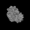
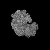
Solanum lycopersicum (tomato)
Method
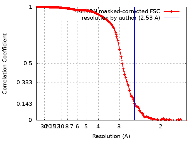
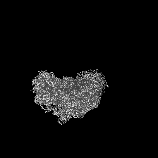
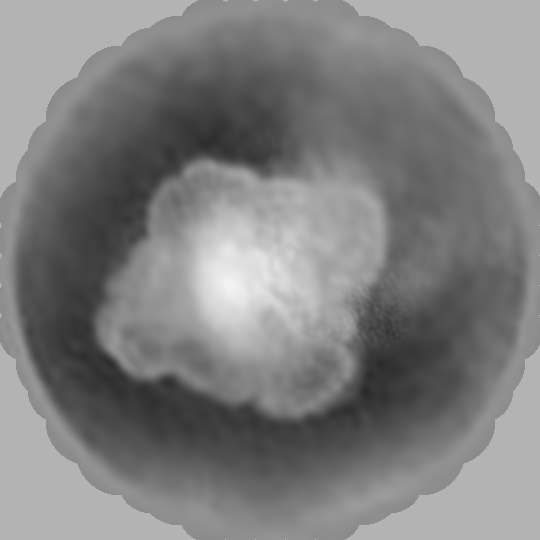
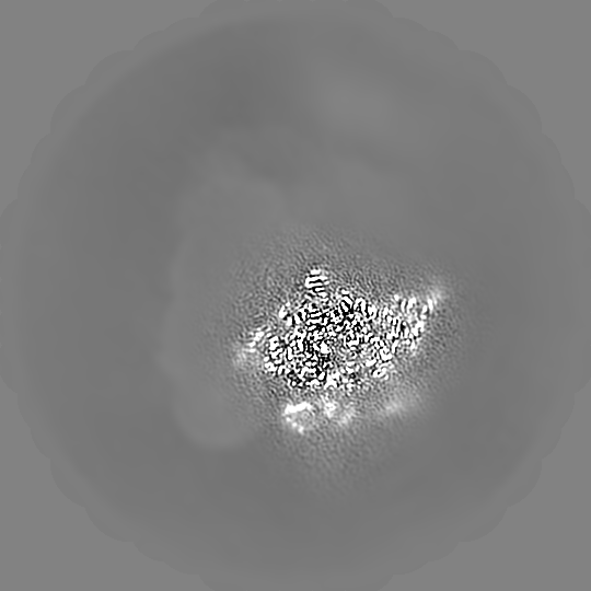
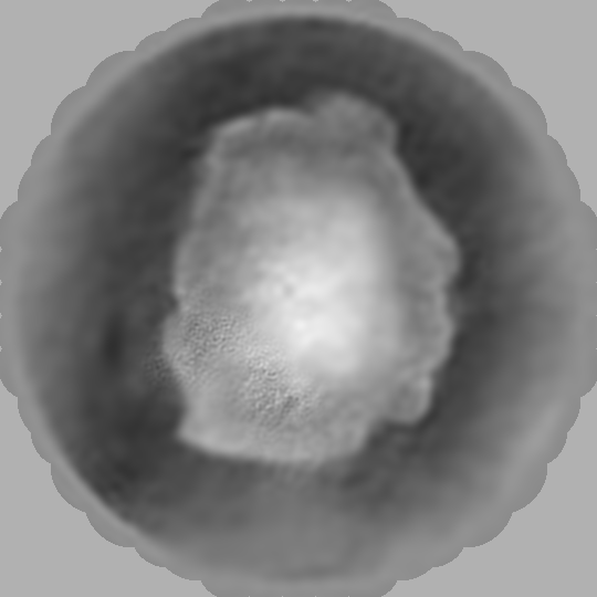
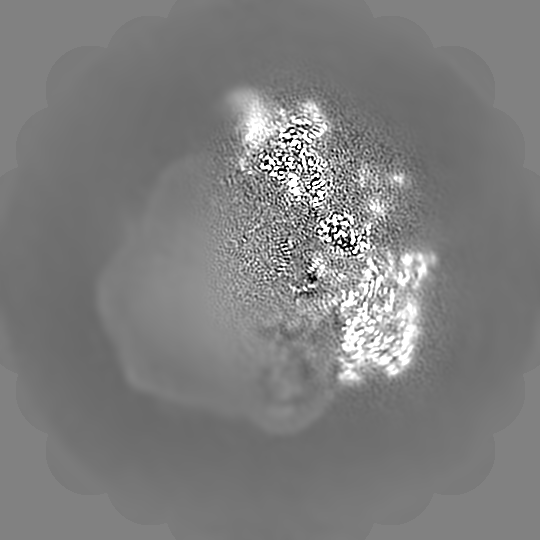
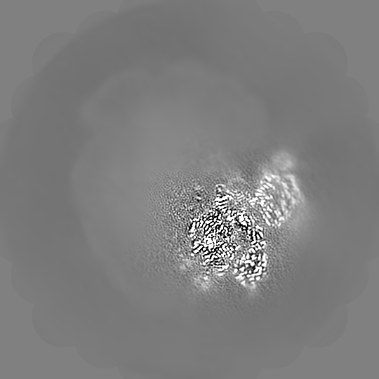
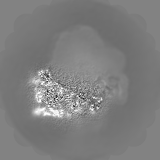
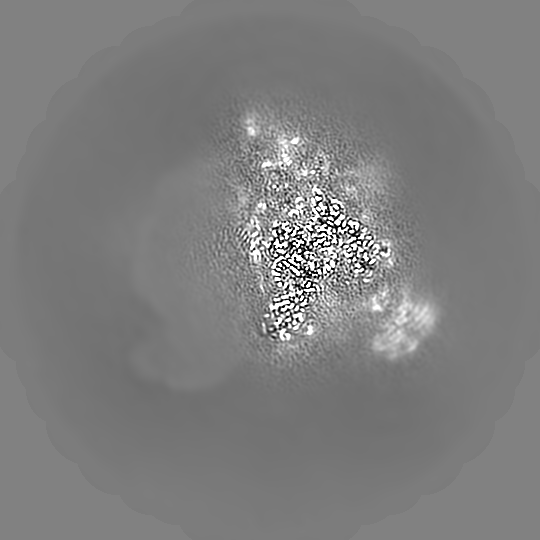
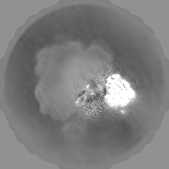
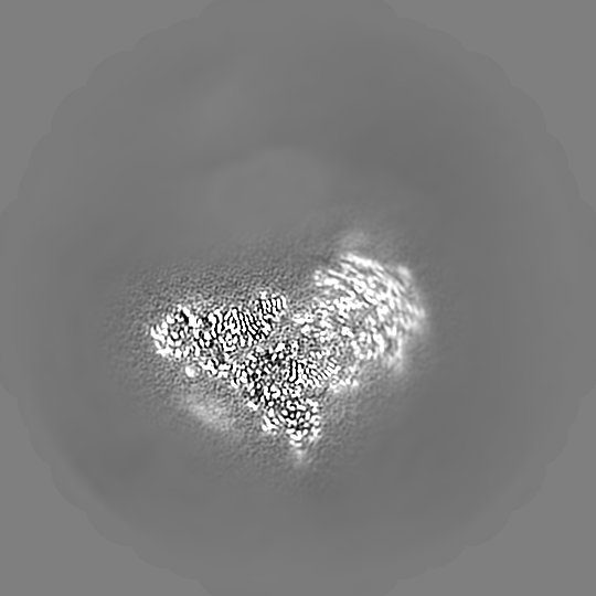
single particle reconstruction / cryo EM / Resolution: 2.53 Å
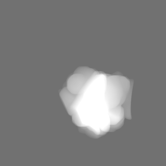
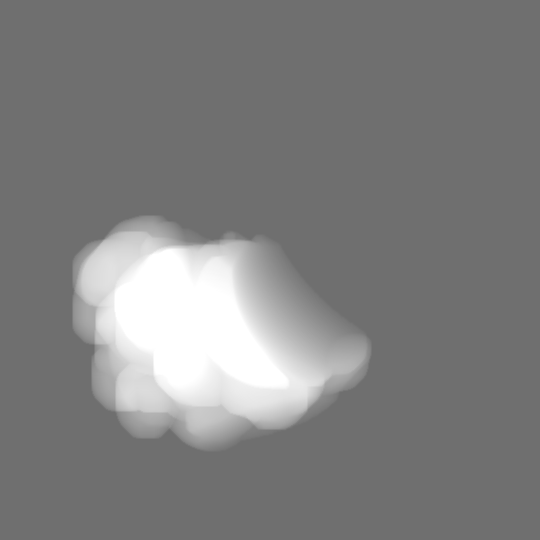
Journal: Plant Commun / Year: 2022 Title: Cryo-EM structure and rRNA modification sites of a plant ribosome. Authors: Patrick Cottilli / Yuzuru Itoh / Yuko Nobe / Anton S Petrov / Purificación Lisón / Masato Taoka / Alexey Amunts / Abstract: Protein synthesis in crop plants contributes to the balance of food and fuel on our planet, which influences human metabolic activity and lifespan. Protein synthesis can be regulated with respect to ...Protein synthesis in crop plants contributes to the balance of food and fuel on our planet, which influences human metabolic activity and lifespan. Protein synthesis can be regulated with respect to changing environmental cues via the deposition of chemical modifications into rRNA. Here, we present the structure of a plant ribosome from tomato and a quantitative mass spectrometry analysis of its rRNAs. The study reveals fine features of the ribosomal proteins and 71 plant-specific rRNA modifications, and it re-annotates 30 rRNA residues in the available sequence. At the protein level, isoAsp is found in position 137 of uS11, and a zinc finger previously believed to be universal is missing from eL34, suggesting a lower effect of zinc deficiency on protein synthesis in plants. At the rRNA level, the plant ribosome differs markedly from its human counterpart with respect to the spatial distribution of modifications. Thus, it represents an additional layer of gene expression regulation, highlighting the molecular signature of a plant ribosome. The results provide a reference model of a plant ribosome for structural studies and an accurate marker for molecular ecology.
In the structure databanks used in Yorodumi, some data are registered as the other names, "COVID-19 virus" and "2019-nCoV". Here are the details of the virus and the list of structure data.
Jan 31, 2019. EMDB accession codes are about to change! (news from PDBe EMDB page)
EMDB accession codes are about to change! (news from PDBe EMDB page)
The allocation of 4 digits for EMDB accession codes will soon come to an end. Whilst these codes will remain in use, new EMDB accession codes will include an additional digit and will expand incrementally as the available range of codes is exhausted. The current 4-digit format prefixed with “EMD-” (i.e. EMD-XXXX) will advance to a 5-digit format (i.e. EMD-XXXXX), and so on. It is currently estimated that the 4-digit codes will be depleted around Spring 2019, at which point the 5-digit format will come into force.
The EM Navigator/Yorodumi systems omit the EMD- prefix.
Related info.:Q: What is EMD? / ID/Accession-code notation in Yorodumi/EM Navigator
Yorodumi is a browser for structure data from EMDB, PDB, SASBDB, etc.
This page is also the successor to EM Navigator detail page, and also detail information page/front-end page for Omokage search.
The word "yorodu" (or yorozu) is an old Japanese word meaning "ten thousand". "mi" (miru) is to see.
Related info.:EMDB / PDB / SASBDB / Comparison of 3 databanks / Yorodumi Search / Aug 31, 2016. New EM Navigator & Yorodumi / Yorodumi Papers / Jmol/JSmol / Function and homology information / Changes in new EM Navigator and Yorodumi
 Movie
Movie Controller

 Yorodumi
 Open data
 Basic information
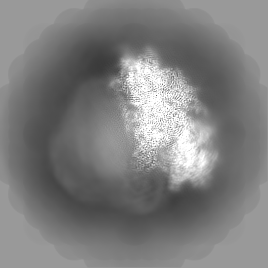
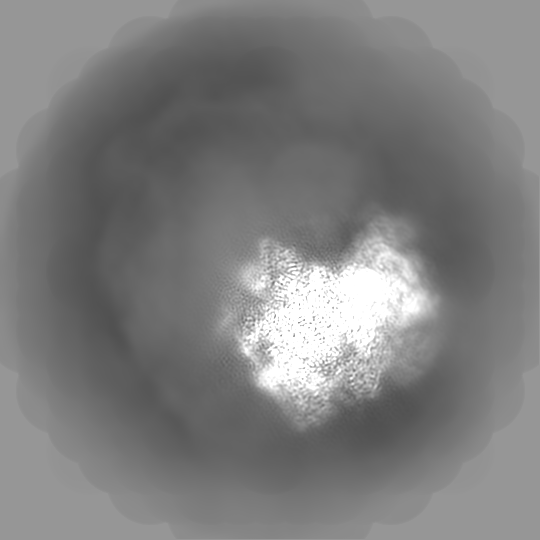
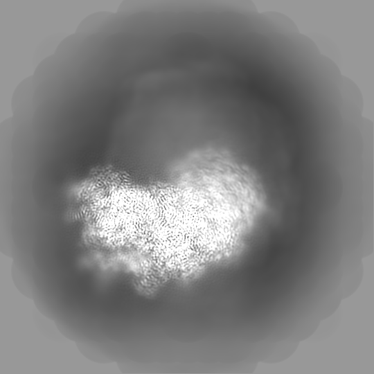
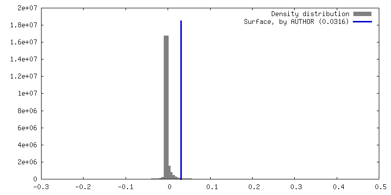
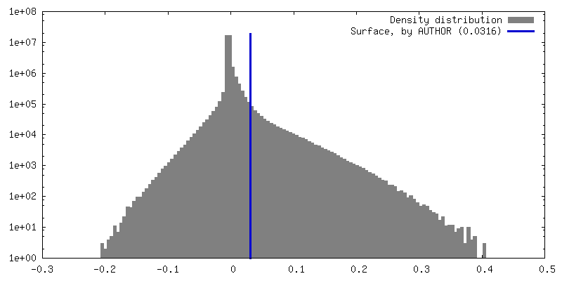
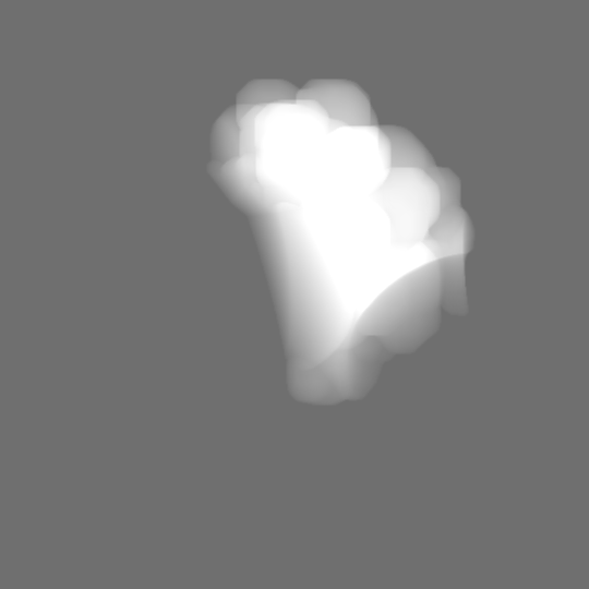
 Map data
 Sample
 Keywords
 Function and homology information
 Authors
 Citation


 Structure visualization
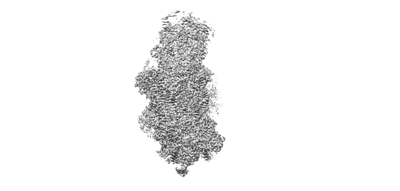
 Downloads & links
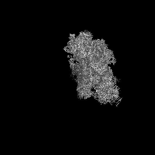
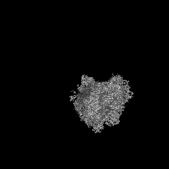
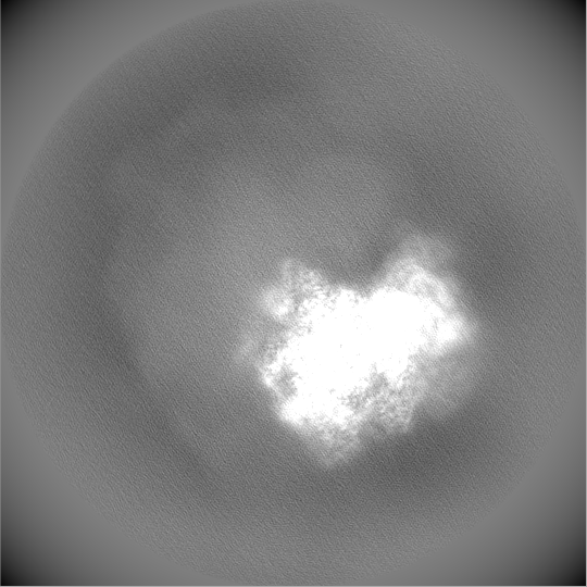
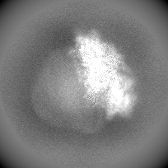
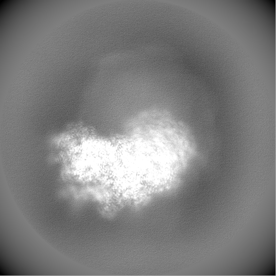
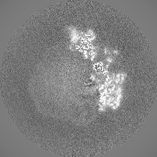
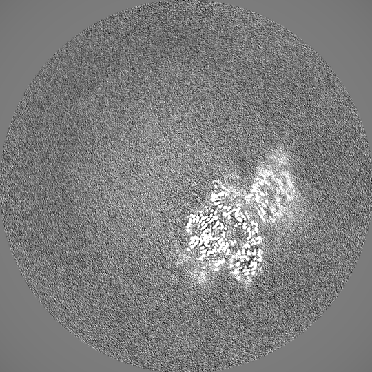
 emd_14002.png
 http://ftp.pdbj.org/pub/emdb/structures/EMD-14002


 Z (Sec.)
 Y (Row.)
 X (Col.)


 Sample components
 Processing
 Electron microscopy
 FIELD EMISSION GUN