ムービー
ムービー コントローラー


+ データを開く

- 基本情報
基本情報
| 登録情報 |  | |||||||||
|---|---|---|---|---|---|---|---|---|---|---|
| タイトル | Fibrillin-1 microfibril arm-region | |||||||||
 マップデータ マップデータ | Fibrillin microfibril arm-region | |||||||||
 試料 試料 |
| |||||||||
 キーワード キーワード | Microfibril / Extra-Cellular-Matrix / STRUCTURAL PROTEIN | |||||||||
| 生物種 |  | |||||||||
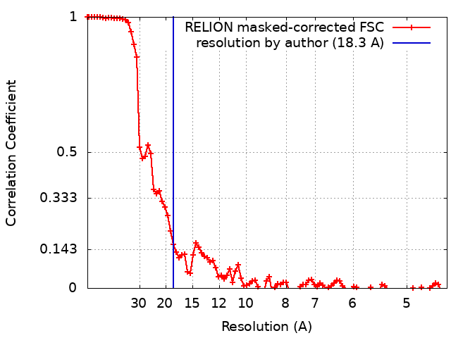
| 手法 | 単粒子再構成法 / クライオ電子顕微鏡法 / 解像度: 18.3 Å | |||||||||
 データ登録者 データ登録者 | Godwin ARF / Thomson J / Holmes DF / Adamo CS / Sengle G / Sherratt MJ / Roseman AM / Baldock C | |||||||||
| 資金援助 |  英国, 2件 英国, 2件
| |||||||||
 引用 引用 | ジャーナル: Nat Struct Mol Biol / 年: 2023 タイトル: Fibrillin microfibril structure identifies long-range effects of inherited pathogenic mutations affecting a key regulatory latent TGFβ-binding site. 著者: Alan R F Godwin / Rana Dajani / Xinyang Zhang / Jennifer Thomson / David F Holmes / Christin S Adamo / Gerhard Sengle / Michael J Sherratt / Alan M Roseman / Clair Baldock /  要旨: Genetic mutations in fibrillin microfibrils cause serious inherited diseases, such as Marfan syndrome and Weill-Marchesani syndrome (WMS). These diseases typically show major dysregulation of tissue ...Genetic mutations in fibrillin microfibrils cause serious inherited diseases, such as Marfan syndrome and Weill-Marchesani syndrome (WMS). These diseases typically show major dysregulation of tissue development and growth, particularly in skeletal long bones, but links between the mutations and the diseases are unknown. Here we describe a detailed structural analysis of native fibrillin microfibrils from mammalian tissue by cryogenic electron microscopy. The major bead region showed pseudo eightfold symmetry where the amino and carboxy termini reside. On the basis of this structure, we show that a WMS deletion mutation leads to the induction of a structural rearrangement that blocks interaction with latent TGFβ-binding protein-1 at a remote site. Separate deletion of this binding site resulted in the assembly of shorter fibrillin microfibrils with structural alterations. The integrin αβ-binding site was also mapped onto the microfibril structure. These results establish that in complex extracellular assemblies, such as fibrillin microfibrils, mutations may have long-range structural consequences leading to the disruption of growth factor signaling and the development of disease. | |||||||||
| 履歴 |
|
- 構造の表示
構造の表示
| 添付画像 |
|---|

- ダウンロードとリンク
-EMDBアーカイブ
| マップデータ | emd_13986.map.gz | 48 MB |  EMDBマップデータ形式 EMDBマップデータ形式 | |
|---|---|---|---|---|
| ヘッダ (付随情報) | emd-13986-v30.xmlemd-13986.xml | 15.2 KB 15.2 KB | 表示 表示 | EMDBヘッダ |
| FSC (解像度算出) | emd_13986_fsc.xml | 9.2 KB | 表示 | FSCデータファイル |
| 画像 |  emd_13986.png emd_13986.png | 23.2 KB | ||
| Filedesc metadata | emd-13986.cif.gz | 4.6 KB | ||
| その他 | emd_13986_additional_1.map.gz | 47.3 MB | ||
| アーカイブディレクトリ |  http://ftp.pdbj.org/pub/emdb/structures/EMD-13986ftp://ftp.pdbj.org/pub/emdb/structures/EMD-13986 http://ftp.pdbj.org/pub/emdb/structures/EMD-13986ftp://ftp.pdbj.org/pub/emdb/structures/EMD-13986 | HTTPS FTP |
-検証レポート
| 文書・要旨 | emd_13986_validation.pdf.gz | 458.1 KB | 表示 | EMDB検証レポート |
|---|---|---|---|---|
| 文書・詳細版 | emd_13986_full_validation.pdf.gz | 457.6 KB | 表示 | |
| XML形式データ | emd_13986_validation.xml.gz | 11 KB | 表示 | |
| CIF形式データ | emd_13986_validation.cif.gz | 14.4 KB | 表示 | |
| アーカイブディレクトリ | https://ftp.pdbj.org/pub/emdb/validation_reports/EMD-13986ftp://ftp.pdbj.org/pub/emdb/validation_reports/EMD-13986 | HTTPS FTP |
-関連構造データ
-リンク
| EMDBのページ | EMDB (EBI/PDBe) / EMDataResource |
|---|
-マップ
| ファイル | ダウンロード / ファイル: emd_13986.map.gz / 形式: CCP4 / 大きさ: 64 MB / タイプ: IMAGE STORED AS FLOATING POINT NUMBER (4 BYTES) | ||||||||||||||||||||
|---|---|---|---|---|---|---|---|---|---|---|---|---|---|---|---|---|---|---|---|---|---|
| 注釈 | Fibrillin microfibril arm-region | ||||||||||||||||||||
| ボクセルのサイズ | X=Y=Z: 2.22 Å | ||||||||||||||||||||
| 密度 |
| ||||||||||||||||||||
| 対称性 | 空間群: 1 | ||||||||||||||||||||
| 詳細 | EMDB XML:
|
-添付データ
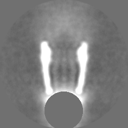
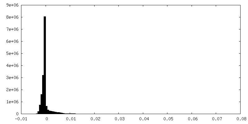
-追加マップ: Fibrillin microfibril arm-region with bread region density removed
| ファイル | emd_13986_additional_1.map | ||||||||||||
|---|---|---|---|---|---|---|---|---|---|---|---|---|---|
| 注釈 | Fibrillin microfibril arm-region with bread region density removed | ||||||||||||
| 投影像・断面図 |
| ||||||||||||
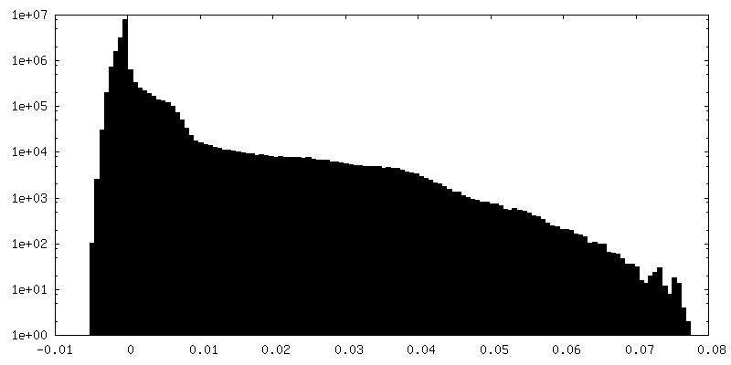
| 密度ヒストグラム |
 Z
Z Y
Y X
X


- 試料の構成要素
-全体 : Fibrillin-1 microfibril arm region
| 全体 | 名称: Fibrillin-1 microfibril arm region |
|---|---|
| 要素 |
|
-超分子 #1: Fibrillin-1 microfibril arm region
| 超分子 | 名称: Fibrillin-1 microfibril arm region / タイプ: tissue / ID: 1 / 親要素: 0 詳細: Microfibrils generated from sonication of bovine ciliary zonule tissue. |
|---|---|
| 由来(天然) | 生物種: |
-実験情報
-構造解析
| 手法 | クライオ電子顕微鏡法 |
|---|---|
 解析 解析 | 単粒子再構成法 |
| 試料の集合状態 | filament |
-試料調製
| 濃度 | 0.1 mg/mL | ||||||||||||
|---|---|---|---|---|---|---|---|---|---|---|---|---|---|
| 緩衝液 | pH: 7.4 構成要素:
詳細: Solutions were made fresh and filtered using 0.2 um filters. | ||||||||||||
| グリッド | モデル: Quantifoil R1.2/1.3 / 材質: COPPER / メッシュ: 300 / 支持フィルム - 材質: CARBON / 支持フィルム - トポロジー: HOLEY ARRAY / 前処理 - タイプ: GLOW DISCHARGE / 前処理 - 時間: 60 sec. | ||||||||||||
| 凍結 | 凍結剤: ETHANE / チャンバー内湿度: 100 % / チャンバー内温度: 277 K / 装置: FEI VITROBOT MARK IV / 詳細: Blot for 4 seconds before plunging. |
- 電子顕微鏡法
| 顕微鏡 | FEI TITAN KRIOS |
|---|---|
| 撮影 | フィルム・検出器のモデル: GATAN K2 SUMMIT (4k x 4k) 検出モード: COUNTING / デジタル化 - サイズ - 横: 4000 pixel / デジタル化 - サイズ - 縦: 4000 pixel / デジタル化 - 画像ごとのフレーム数: 1-20 / 撮影したグリッド数: 1 / 平均露光時間: 20.0 sec. / 平均電子線量: 66.0 e/Å2 / 詳細: Movies were collected in counting mode |
| 電子線 | 加速電圧: 300 kV / 電子線源:  FIELD EMISSION GUN FIELD EMISSION GUN |
| 電子光学系 | C2レンズ絞り径: 100.0 µm / 照射モード: FLOOD BEAM / 撮影モード: BRIGHT FIELD / Cs: 2.7 mm / 最大 デフォーカス(公称値): 5.0 µm / 最小 デフォーカス(公称値): 2.0 µm / 倍率(公称値): 64000 |
| 試料ステージ | 試料ホルダーモデル: FEI TITAN KRIOS AUTOGRID HOLDER ホルダー冷却材: NITROGEN |
| 実験機器 |  モデル: Titan Krios / 画像提供: FEI Company |