maturation of LSU-rRNA from tricistronic rRNA transcript (SSU-rRNA, 5.8S rRNA, LSU-rRNA) / maturation of SSU-rRNA from tricistronic rRNA transcript (SSU-rRNA, 5.8S rRNA, LSU-rRNA) / maturation of SSU-rRNA / rRNA processing / large ribosomal subunit / ribosomal small subunit biogenesis / ribosomal small subunit assembly / small ribosomal subunit / 5S rRNA binding / small ribosomal subunit rRNA binding ...maturation of LSU-rRNA from tricistronic rRNA transcript (SSU-rRNA, 5.8S rRNA, LSU-rRNA) / maturation of SSU-rRNA from tricistronic rRNA transcript (SSU-rRNA, 5.8S rRNA, LSU-rRNA) / maturation of SSU-rRNA / rRNA processing / large ribosomal subunit / ribosomal small subunit biogenesis / ribosomal small subunit assembly / small ribosomal subunit / 5S rRNA binding / small ribosomal subunit rRNA binding / ribosomal large subunit assembly / cytosolic small ribosomal subunit / large ribosomal subunit rRNA binding / cytosolic large ribosomal subunit / cytoplasmic translation / negative regulation of translation / rRNA binding / structural constituent of ribosome / ribosome / translation / ribonucleoprotein complex / mRNA binding / nucleolus / RNA binding / zinc ion binding / nucleus / cytosol / cytoplasm Similarity search - Function
CXXC motif containing zinc binding protein, eukaryotic / CXXC motif containing zinc binding protein, eukaryotic / Ribosomal L15/L27a, N-terminal / Ribosomal protein L13e / : / Ribosomal protein L13e / Ribosomal protein L30/YlxQ / Ribosomal protein S26e / Ribosomal protein S26e superfamily / Ribosomal protein S26e ...CXXC motif containing zinc binding protein, eukaryotic / CXXC motif containing zinc binding protein, eukaryotic / Ribosomal L15/L27a, N-terminal / Ribosomal protein L13e / : / Ribosomal protein L13e / Ribosomal protein L30/YlxQ / Ribosomal protein S26e / Ribosomal protein S26e superfamily / Ribosomal protein S26e / Ribosomal protein L1 / Ribosomal protein L29e / Ribosomal L29e protein family / Ribosomal protein S5, eukaryotic/archaeal / Ribosomal protein S21e / Ribosomal protein S21e superfamily / Ribosomal protein S21e / Ribosomal protein S2, eukaryotic / Ribosomal protein L22e / Ribosomal protein L22e superfamily / Ribosomal L22e protein family / Ribosomal protein L10e, conserved site / Ribosomal protein L10e signature. / S27a-like superfamily / Ribosomal protein L10e / Ribosomal protein L1, 3-layer alpha/beta-sandwich / Ribosomal protein L19, eukaryotic / Plectin/S10, N-terminal / Plectin/S10 domain / 60S ribosomal protein L18a/ L20, eukaryotes / : / Ribosomal protein S25 / S25 ribosomal protein / Ribosomal protein L44e / Ribosomal protein L44 / Ribosomal protein L34e, conserved site / Ribosomal protein L34e signature. / Ribosomal protein S2, eukaryotic/archaeal / Ribosomal protein L5 eukaryotic, C-terminal / 50S ribosomal protein L18Ae/60S ribosomal protein L20 and L18a / Ribosomal L18 C-terminal region / Ribosomal L40e family / Ribosomal protein 50S-L18Ae/60S-L20/60S-L18A / Ribosomal proteins 50S-L18Ae/60S-L20/60S-L18A / Ribosomal protein 60S L18 and 50S L18e / Ribosomal_L40e / Ribosomal protein L40e / Ribosomal protein L40e superfamily / Ribosomal protein S3, eukaryotic/archaeal / Eukaryotic Ribosomal Protein L27, KOW domain / Ribosomal Protein L6, KOW domain / Ribosomal protein L27e / Ribosomal protein L27e superfamily / Ribosomal L27e protein family / Ribosomal protein S19e / Ribosomal protein S19e / Ribosomal_S19e / Ribosomal protein S6, eukaryotic / Ribosomal protein L39e, conserved site / Ribosomal protein L39e signature. / Ribosomal protein L1-like / Ribosomal protein L1/ribosomal biogenesis protein / 60S ribosomal protein L19 / Ribosomal protein L1p/L10e family / 40S ribosomal protein S1/3, eukaryotes / : / Ribosomal protein L13, eukaryotic/archaeal / Ribosomal protein S19A/S15e / Ribosomal protein L34Ae / Ribosomal protein L34e / 40S ribosomal protein S11, N-terminal / Ribosomal_S17 N-terminal / Ribosomal protein S17e / Ribosomal protein S17e-like superfamily / Ribosomal S17 / Ribosomal protein L7A/L8 / 60S ribosomal protein L6E / 60S ribosomal protein L35 / : / Ribosomal protein L18e / Ribosomal protein S4e, N-terminal / RS4NT (NUC023) domain / Ribosomal protein L19e, C-terminal domain / Ribosomal_L19e / Ribosomal protein L19/L19e / Ribosomal protein L19/L19e, domain 1 / Ribosomal protein L19/L19e superfamily / Ribosomal protein L19e, N-terminal domain / Ribosomal protein L37ae / Ribosomal L37ae protein family / Ribosomal protein L36e / Ribosomal protein L36e domain superfamily / Ribosomal protein L36e / Ribosomal protein S4e / Ribosomal protein L35A / Ribosomal protein S4e, central region / Ribosomal protein S4e, central domain superfamily / Ribosomal family S4e / Ribosomal protein L35Ae / Ribosomal protein L39e Similarity search - Domain/homology
ECU06_1135 protein / 60S ribosomal protein L29 / ECU06_1215 protein / ECU11_0225 protein / Guanine nucleotide binding protein beta subunit / Large ribosomal subunit protein uL15 / Large ribosomal subunit protein uL3 / 40S RIBOSOMAL PROTEIN S15A (S22 in yeast) / 40S RIBOSOMAL PROTEIN S28 / Small ribosomal subunit protein uS3 ...ECU06_1135 protein / 60S ribosomal protein L29 / ECU06_1215 protein / ECU11_0225 protein / Guanine nucleotide binding protein beta subunit / Large ribosomal subunit protein uL15 / Large ribosomal subunit protein uL3 / 40S RIBOSOMAL PROTEIN S15A (S22 in yeast) / 40S RIBOSOMAL PROTEIN S28 / Small ribosomal subunit protein uS3 / Large ribosomal subunit protein eL39 / Small ribosomal subunit protein eS19 / Ribosomal protein L15 / Small ribosomal subunit protein eS7 / 40S RIBOSOMAL PROTEIN S20 / 40S RIBOSOMAL PROTEIN S24 / Large ribosomal subunit protein eL42 / Small ribosomal subunit protein uS12 / 60S RIBOSOMAL PROTEIN L23A / 60S RIBOSOMAL PROTEIN L6 / Large ribosomal subunit protein uL16 / Large ribosomal subunit protein uL14 / Small ribosomal subunit protein uS15 / Small ribosomal subunit protein eS4 / Large ribosomal subunit protein uL4 / 60S ribosomal protein L20 / 60S RIBOSOMAL PROTEIN L26 / RIBOSOMAL PROTEIN S15 / Small ribosomal subunit protein uS5 / Large ribosomal subunit protein eL37 / 60S RIBOSOMAL PROTEIN L17 / UBIQUITIN/ L40 RIBOSOMAL PROTEIN FUSION / 60S RIBOSOMAL PROTEIN L37A (L43) / Small ribosomal subunit protein eS26 / Large ribosomal subunit protein eL36 / Small ribosomal subunit protein uS13 / 60S RIBOSOMAL PROTEIN L19 / 60S RIBOSOMAL PROTEIN L5 / 60S RIBOSOMAL PROTEIN L30 / Small ribosomal subunit protein uS4 / Large ribosomal subunit protein eL21 / Small ribosomal subunit protein eS6 / Large ribosomal subunit protein uL1 / Small ribosomal subunit protein eS1 / 60S RIBOSOMAL PROTEIN L13A (L16) / 40S RIBOSOMAL PROTEIN S10 / Large ribosomal subunit protein eL32 / 40S RIBOSOMAL PROTEIN S27 / Large ribosomal subunit protein eL22 / Small ribosomal subunit protein uS17 / Small ribosomal subunit protein uS2 / 60S RIBOSOMAL PROTEIN L27 / Small ribosomal subunit protein uS7 / 60S RIBOSOMAL PROTEIN L18 / Large ribosomal subunit protein uL30 / Large ribosomal subunit protein eL34 / Small ribosomal subunit protein uS11 / 60S RIBOSOMAL PROTEIN L13 / 40S RIBOSOMAL PROTEIN S16 / Large ribosomal subunit protein eL31 / 60S RIBOSOMAL PROTEIN L35A (L33) / 60S RIBOSOMAL PROTEIN L9 / Small ribosomal subunit protein eS17 / Large ribosomal subunit protein eL8 / Large ribosomal subunit protein uL5 / 40S RIBOSOMAL PROTEIN S12 / Large ribosomal subunit protein uL2 / Large ribosomal subunit protein uL29A / Small ribosomal subunit protein eS25 / Similarity to 60S RIBOSOMAL PROTEIN L24 / Similarity to monoubiquitin/carboxy-extension protein fusion / 40S ribosomal protein S8 / Uncharacterized protein ECU01_0250 Similarity search - Component
H2020 Marie Curie Actions of the European Commission
97617
United Kingdom
H2020 Marie Curie Actions of the European Commission
895166
United Kingdom
Wellcome Trust
108466/Z/15/Z
United Kingdom
Citation
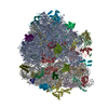
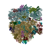
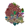
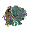
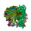
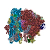
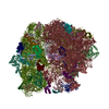
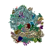
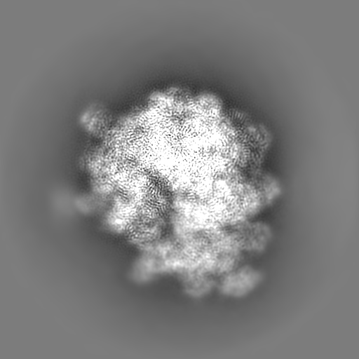
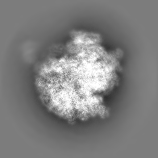
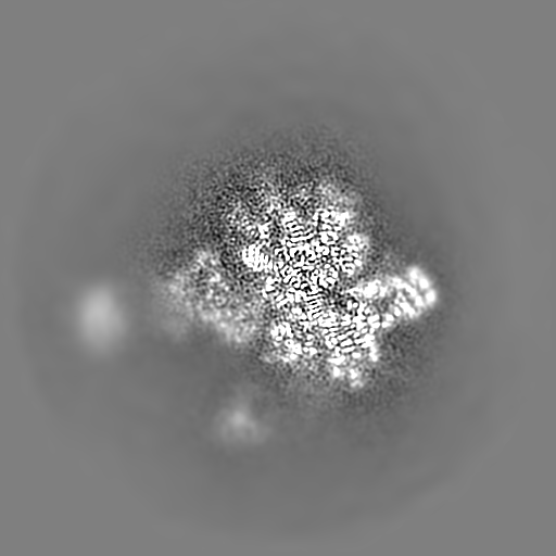
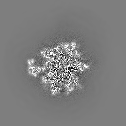
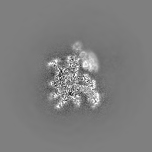
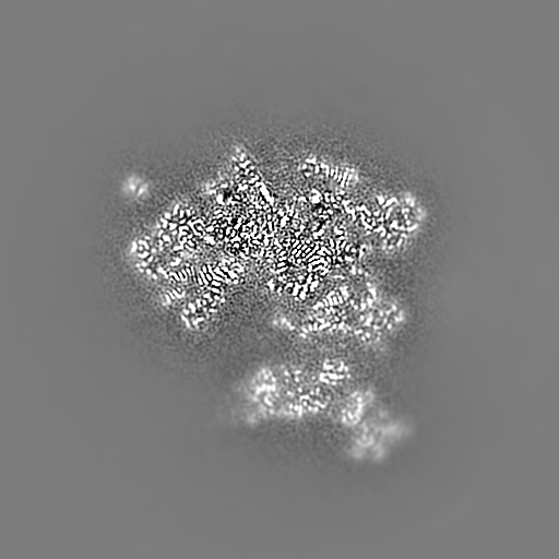
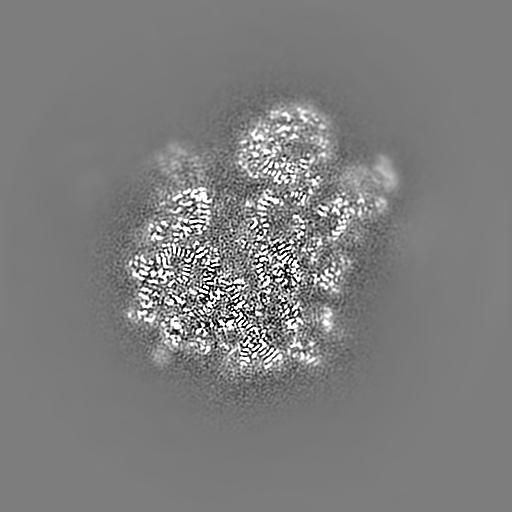
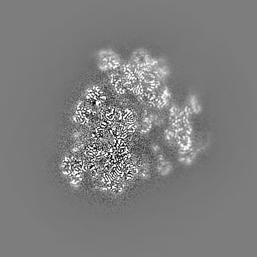
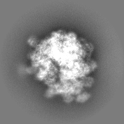
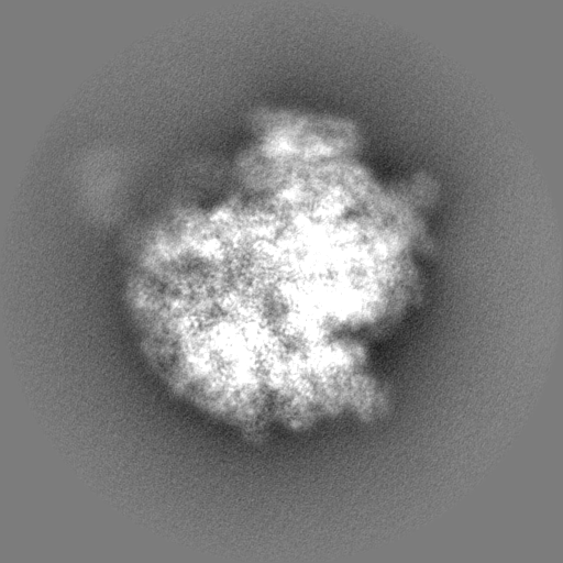
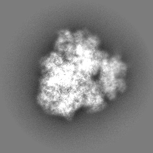
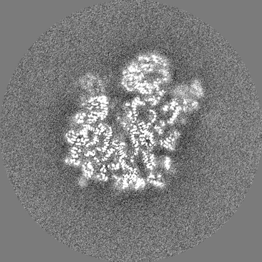
Journal: Nat Commun / Year: 2022 Title: Adaptation to genome decay in the structure of the smallest eukaryotic ribosome. Authors: David Nicholson / Marco Salamina / Johan Panek / Karla Helena-Bueno / Charlotte R Brown / Robert P Hirt / Neil A Ranson / Sergey V Melnikov / Abstract: The evolution of microbial parasites involves the counterplay between natural selection forcing parasites to improve and genetic drifts forcing parasites to lose genes and accumulate deleterious ...The evolution of microbial parasites involves the counterplay between natural selection forcing parasites to improve and genetic drifts forcing parasites to lose genes and accumulate deleterious mutations. Here, to understand how this counterplay occurs at the scale of individual macromolecules, we describe cryo-EM structure of ribosomes from Encephalitozoon cuniculi, a eukaryote with one of the smallest genomes in nature. The extreme rRNA reduction in E. cuniculi ribosomes is accompanied with unparalleled structural changes, such as the evolution of previously unknown molten rRNA linkers and bulgeless rRNA. Furthermore, E. cuniculi ribosomes withstand the loss of rRNA and protein segments by evolving an ability to use small molecules as structural mimics of degenerated rRNA and protein segments. Overall, we show that the molecular structures long viewed as reduced, degenerated, and suffering from debilitating mutations possess an array of compensatory mechanisms that allow them to remain active despite the extreme molecular reduction.
History
Deposition
Dec 3, 2021
-
Header (metadata) release
Feb 9, 2022
-
Map release
Feb 9, 2022
-
Update
Nov 6, 2024
-
Current status
Nov 6, 2024
Processing site: PDBe / Status: Released
-
Structure visualization
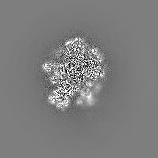
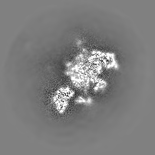
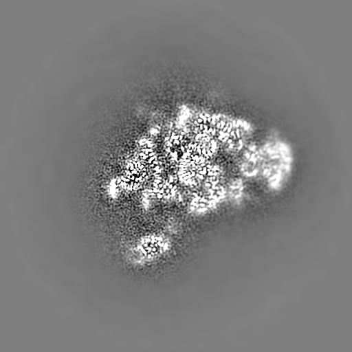
Movie
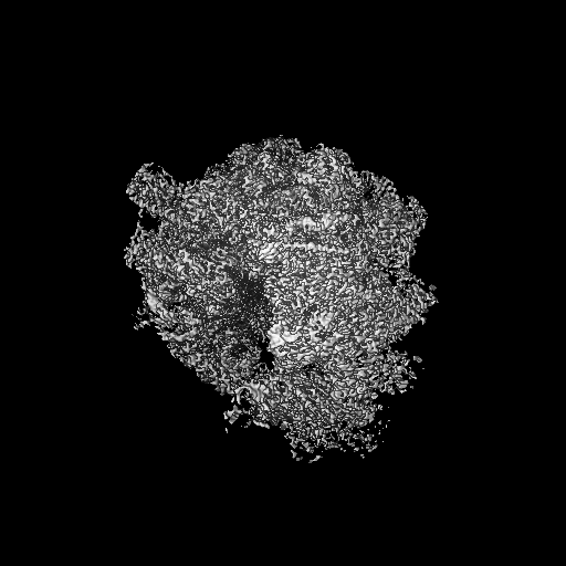
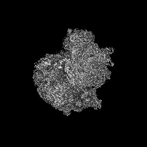
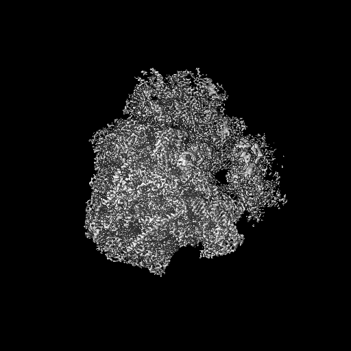
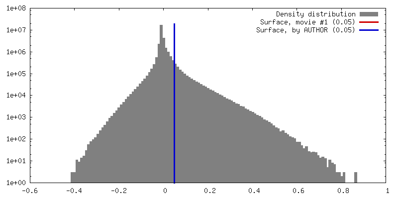
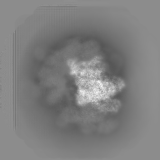
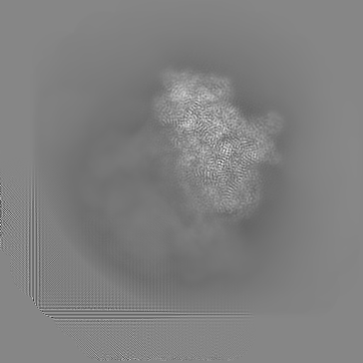
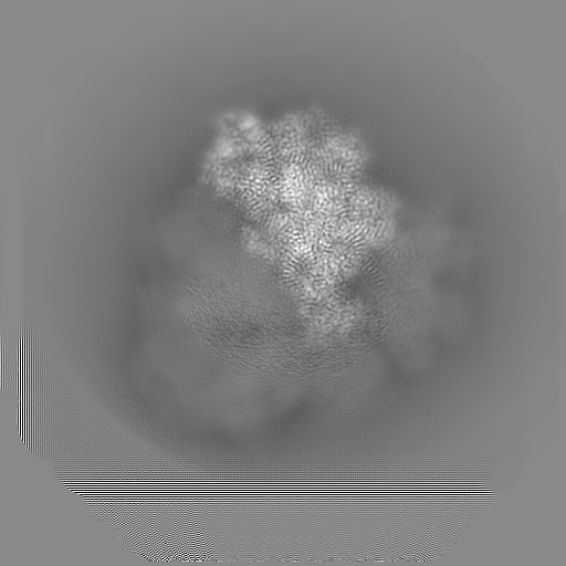
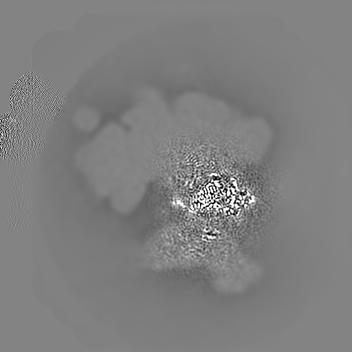
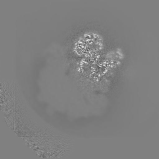
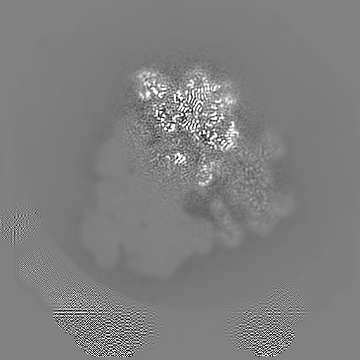
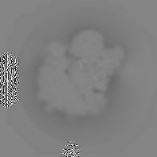
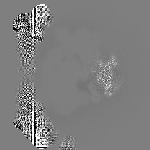
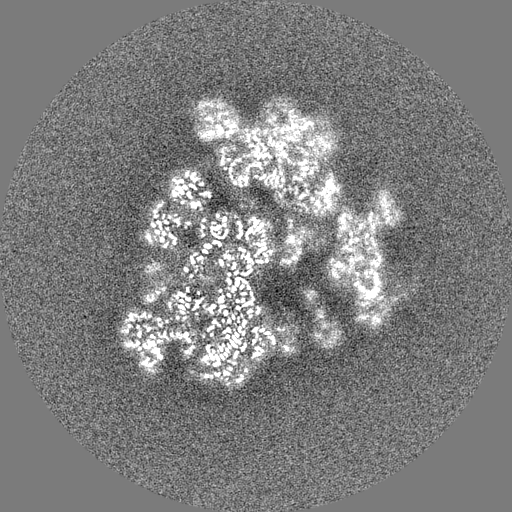
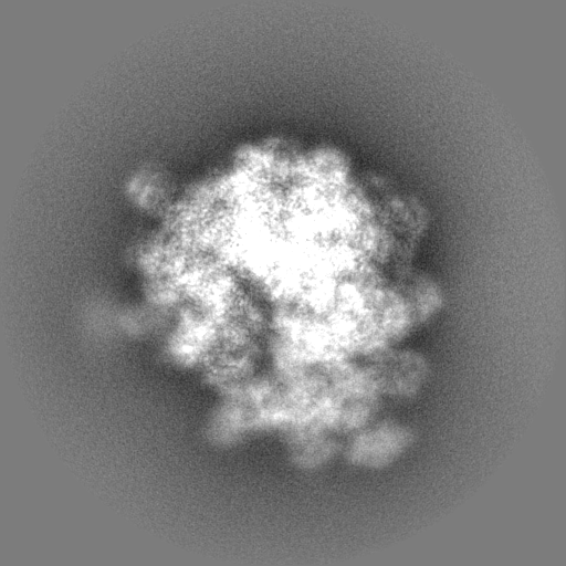
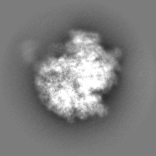
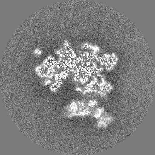
Surface view with section colored by density value
Model: Quantifoil R1.2/1.3 / Material: COPPER / Mesh: 400 / Pretreatment - Type: GLOW DISCHARGE / Pretreatment - Time: 30 sec. / Pretreatment - Atmosphere: AIR / Details: Quorum GloQube, 10 mA
Vitrification
Cryogen name: ETHANE / Chamber humidity: 100 % / Chamber temperature: 277 K / Instrument: FEI VITROBOT MARK IV Details: Quantifoil grids (R1.2/1.3, 400 mesh, copper) were glow discharged (10 mA, 30s, Quorum GloQube), and 3 microlitres of the crude sample of E. cuniculi ribosomes (300 nM) was pipetted onto a ...Details: Quantifoil grids (R1.2/1.3, 400 mesh, copper) were glow discharged (10 mA, 30s, Quorum GloQube), and 3 microlitres of the crude sample of E. cuniculi ribosomes (300 nM) was pipetted onto a grid. Excess sample was immediately blotted off and vitrification was performed by plunging the grid into liquid nitrogen-cooled liquid ethane at 100% humidity and 4 degrees celsius using an FEI Vitrobot Mark IV (Thermo Fisher).
-
Electron microscopy
Microscope
FEI TITAN KRIOS
Image recording
Film or detector model: FEI FALCON III (4k x 4k) / Detector mode: INTEGRATING / Digitization - Dimensions - Width: 4096 pixel / Digitization - Dimensions - Height: 4096 pixel / Number real images: 2210 / Average exposure time: 1.35 sec. / Average electron dose: 60.0 e/Å2
Electron beam
Acceleration voltage: 300 kV / Electron source: FIELD EMISSION GUN
Drift-corrected and dose-corrected averages of each movie were created using RELION 3.1s own implementation of motion correction and the contrast transfer functions estimated using CTFFIND-4.1
Particle selection
Number selected: 260895 Details: Particles were picked using Laplacian-of-Gaussian autopicking and reference-free 2D classification used to generate templates for further autopicking. The resulting particles were extracted ...Details: Particles were picked using Laplacian-of-Gaussian autopicking and reference-free 2D classification used to generate templates for further autopicking. The resulting particles were extracted with binning-by-4, and 2D and 3D classification performed to remove junk images
Startup model
Type of model: INSILICO MODEL In silico model: The 3D classification step was carried out using a 60 Angstroms low-passed filtered ab initio starting model made using a stochastic gradient descent procedure.
Final reconstruction
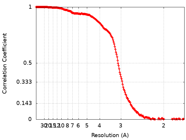
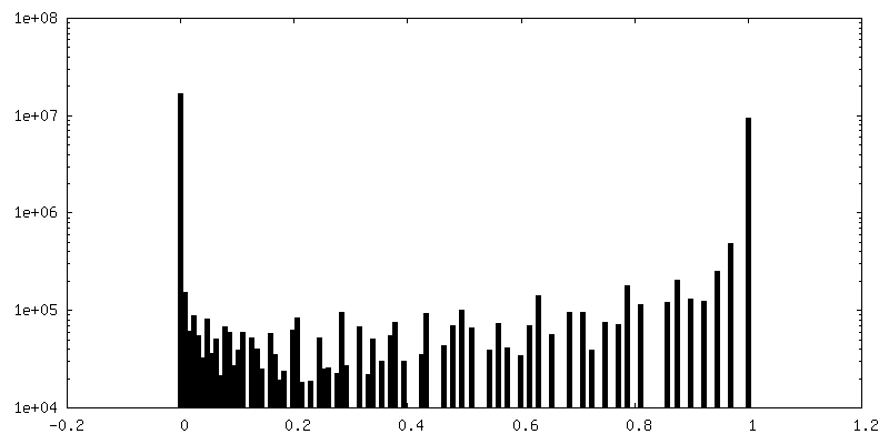
Number classes used: 1 / Applied symmetry - Point group: C1 (asymmetric) / Resolution.type: BY AUTHOR / Resolution: 2.7 Å / Resolution method: FSC 0.143 CUT-OFF / Software - Name: RELION (ver. 3.1) Details: The remaining particles were re-extracted without binning and aligned and refined in 3D, again using a 60 Angstrom low-passed filtered ab initio starting model. Rounds of CTF refinement and ...Details: The remaining particles were re-extracted without binning and aligned and refined in 3D, again using a 60 Angstrom low-passed filtered ab initio starting model. Rounds of CTF refinement and Bayesian polishing were performed until the map resolution stopped improving. 108,005 particles fed into the final 3D reconstruction of estimated resolution 2.7 Angstrom. Number images used: 108005
Initial angle assignment
Type: MAXIMUM LIKELIHOOD / Software - Name: RELION (ver. 3.1)
Final angle assignment
Type: MAXIMUM LIKELIHOOD / Software - Name: RELION (ver. 3.1)
source_name: PDB, initial_model_type: experimental model
Details
The model was built using fragments of S. cerevisiae (pdb id 4v88) and V. necatrix ribosomes (pdb id 6rm3) as starting models that were edited using Coot using genomic sequences of the E. cuniculi strain GB-M1 to model rRNA and ribosomal proteins. For ribosomal proteins that are encoded by two alternative genes (with one gene coding for a zinc-coordinating protein and another gene coding for a zinc-free ribosomal protein), we used zinc-coordinating isoforms, because the cryo-EM map revealed the presence of these isoforms and not their zinc-free paralogs in the ribosome structure. The identity of protein msL2 in the ribosome structure was determined using the genomic sequence of the E. cuniculi strain GB-M1 and the cryo-EM map that revealed a unique combination of aromatic and bulky amino acids in its structure: the cryo-EM map showed that msL2 has a tyrosine residue at position 5, a tryptophan residue at position 9, and lysine or arginine residues at positions 10, 12 and 13. The only protein with this sequence was the hypothetical protein ECU06_1135, whose sequence and length were fully consistent with the cryo-EM map. The structure of E. cuniculi ribosomes was refined using Phenix real space refine and validated using MolProbity within Phenix and PDB OneDep. The parts of the model corresponding to the 60S, 40S body and 40S head were built and refined using the consensus map, 40S body multibody map and 40S head multibody map, respectively.
Refinement
Space: REAL / Protocol: OTHER / Target criteria: correlation coefficient
Output model
PDB-7qep: Cryo-EM structure of the ribosome from Encephalitozoon cuniculi
+
About Yorodumi
-
News
-
Feb 9, 2022. New format data for meta-information of EMDB entries
New format data for meta-information of EMDB entries
Version 3 of the EMDB header file is now the official format.
The previous official version 1.9 will be removed from the archive.
In the structure databanks used in Yorodumi, some data are registered as the other names, "COVID-19 virus" and "2019-nCoV". Here are the details of the virus and the list of structure data.
Jan 31, 2019. EMDB accession codes are about to change! (news from PDBe EMDB page)
EMDB accession codes are about to change! (news from PDBe EMDB page)
The allocation of 4 digits for EMDB accession codes will soon come to an end. Whilst these codes will remain in use, new EMDB accession codes will include an additional digit and will expand incrementally as the available range of codes is exhausted. The current 4-digit format prefixed with “EMD-” (i.e. EMD-XXXX) will advance to a 5-digit format (i.e. EMD-XXXXX), and so on. It is currently estimated that the 4-digit codes will be depleted around Spring 2019, at which point the 5-digit format will come into force.
The EM Navigator/Yorodumi systems omit the EMD- prefix.
Related info.:Q: What is EMD? / ID/Accession-code notation in Yorodumi/EM Navigator
Yorodumi is a browser for structure data from EMDB, PDB, SASBDB, etc.
This page is also the successor to EM Navigator detail page, and also detail information page/front-end page for Omokage search.
The word "yorodu" (or yorozu) is an old Japanese word meaning "ten thousand". "mi" (miru) is to see.
Related info.:EMDB / PDB / SASBDB / Comparison of 3 databanks / Yorodumi Search / Aug 31, 2016. New EM Navigator & Yorodumi / Yorodumi Papers / Jmol/JSmol / Function and homology information / Changes in new EM Navigator and Yorodumi
 Movie
Movie Controller

 Yorodumi
 Open data
 Basic information
 Map data
 Sample
 Keywords
 Function and homology information
 Encephalitozoon cuniculi (fungus) /
  Authors
 United Kingdom, 6 items
  Citation
 Structure visualization
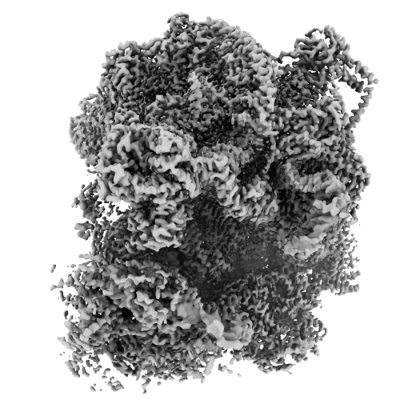
 Downloads & links
 emd_13936.png
 http://ftp.pdbj.org/pub/emdb/structures/EMD-13936


 Z (Sec.)
 Y (Row.)
 X (Col.)


 Sample components

 Processing
 Electron microscopy
 FIELD EMISSION GUN