ribosomal subunit / preribosome / nonfunctional rRNA decay / preribosome, small subunit precursor / translational elongation / ribosomal subunit export from nucleus / translational termination / translation regulator activity / ribosomal small subunit export from nucleus / DNA-(apurinic or apyrimidinic site) endonuclease activity ...ribosomal subunit / preribosome / nonfunctional rRNA decay / preribosome, small subunit precursor / translational elongation / ribosomal subunit export from nucleus / translational termination / translation regulator activity / ribosomal small subunit export from nucleus / DNA-(apurinic or apyrimidinic site) endonuclease activity / cytosolic ribosome / maturation of LSU-rRNA from tricistronic rRNA transcript (SSU-rRNA, 5.8S rRNA, LSU-rRNA) / maturation of SSU-rRNA from tricistronic rRNA transcript (SSU-rRNA, 5.8S rRNA, LSU-rRNA) / maturation of SSU-rRNA / maintenance of translational fidelity / protein tag activity / rRNA processing / large ribosomal subunit / ribosomal small subunit assembly / ribosome binding / ribosomal small subunit biogenesis / 5S rRNA binding / ribosomal large subunit assembly / small ribosomal subunit / small ribosomal subunit rRNA binding / large ribosomal subunit rRNA binding / cytosolic small ribosomal subunit / cytosolic large ribosomal subunit / cytoplasmic translation / negative regulation of translation / rRNA binding / structural constituent of ribosome / ribosome / translation / ribonucleoprotein complex / mRNA binding / nucleolus / RNA binding / zinc ion binding / nucleus / cytosol / cytoplasm Similarity search - Function
Ribosomal protein L32e, archaeal / Ribosomal protein S26e / Ribosomal protein S26e superfamily / Ribosomal protein S26e / Small (40S) ribosomal subunit Asc1/RACK1 / Ribosomal protein L22e / Ribosomal protein L22e superfamily / Ribosomal L22e protein family / Ribosomal protein L13e / Ribosomal protein L13e ...Ribosomal protein L32e, archaeal / Ribosomal protein S26e / Ribosomal protein S26e superfamily / Ribosomal protein S26e / Small (40S) ribosomal subunit Asc1/RACK1 / Ribosomal protein L22e / Ribosomal protein L22e superfamily / Ribosomal L22e protein family / Ribosomal protein L13e / Ribosomal protein L13e / Ribosomal protein S5, eukaryotic/archaeal / Ribosomal protein S21e / Ribosomal protein S21e superfamily / Ribosomal protein S21e / 60S ribosomal protein L18a/ L20, eukaryotes / Ribosomal protein L10e, conserved site / Ribosomal protein L10e signature. / 40S Ribosomal protein S10 / Ribosomal protein L44e signature. / S27a-like superfamily / Plectin/S10, N-terminal / Plectin/S10 domain / Ribosomal protein L10e / Ribosomal protein L34e, conserved site / Ribosomal protein L34e signature. / Ribosomal protein L5 eukaryotic, C-terminal / Ribosomal L18 C-terminal region / Ribosomal L40e family / 50S ribosomal protein L18Ae/60S ribosomal protein L20 and L18a / : / Ribosomal protein S25 / Ribosomal protein 50S-L18Ae/60S-L20/60S-L18A / Ribosomal proteins 50S-L18Ae/60S-L20/60S-L18A / S25 ribosomal protein / Ribosomal_L40e / Ribosomal protein L27e / Ribosomal protein L40e / Ribosomal protein L40e superfamily / Ribosomal protein L27e superfamily / Ribosomal L27e protein family / Ribosomal protein L44e / Ribosomal protein L44 / Ribosomal protein 60S L18 and 50S L18e / Ribosomal protein S27a / Ribosomal protein S27a / Ribosomal protein S27a / Ribosomal protein S2, eukaryotic/archaeal / : / Ribosomal protein L7A/L8 / Ribosomal protein L34Ae / 40S ribosomal protein S29/30S ribosomal protein S14 type Z / Ribosomal protein L34e / 60S ribosomal protein L6E / Ribosomal protein L30/YlxQ / 60S ribosomal protein L19 / Ribosomal protein L13, eukaryotic/archaeal / : / Ribosomal protein L36e / Ribosomal protein L36e domain superfamily / Ribosomal protein L36e / 40S ribosomal protein S4, C-terminal domain / 40S ribosomal protein S4 C-terminus / Ribosomal protein S19A/S15e / Ribosomal protein L18e / Ribosomal protein S19e / Ribosomal protein L35A / Ribosomal protein S19e / Ribosomal protein L35Ae / Ribosomal_S19e / Ribosomal protein S4e, N-terminal, conserved site / Ribosomal protein S4e signature. / Ribosomal protein L35A superfamily / : / : / Ribosomal protein L19e, C-terminal domain / Ribosomal_L19e / Ribosomal protein L19/L19e / Ribosomal protein L19/L19e, domain 1 / Ribosomal protein L19/L19e superfamily / Ribosomal protein L19e, N-terminal domain / Ribosomal protein L32e, conserved site / Ribosomal protein L32e signature. / Ribosomal protein L39e / Ribosomal protein L39e domain superfamily / Ribosomal L39 protein / Ribosomal protein L5 eukaryotic/L18 archaeal / Ribosomal large subunit proteins 60S L5, and 50S L18 / 40S ribosomal protein S11, N-terminal / Ribosomal_S17 N-terminal / Ribosomal protein S7e / Ribosomal protein S7e / : / Ribosomal protein S4e, N-terminal / RS4NT (NUC023) domain / Ribosomal protein S4, KOW domain / Ribosomal protein S4e / Ribosomal protein S4e, central region / Ribosomal protein S4e, central domain superfamily / Ribosomal family S4e / Ribosomal protein L31e Similarity search - Domain/homology
40S ribosomal protein S5 / 60S ribosomal protein L6 / 60S ribosomal protein L35a / Small ribosomal subunit protein uS11 / Ribosomal protein L15 / Ribosomal protein L34 / 40S ribosomal protein S12 / 40S Ribosomal protein S19 / Small ribosomal subunit protein uS5 / 60S ribosomal protein L26 ...40S ribosomal protein S5 / 60S ribosomal protein L6 / 60S ribosomal protein L35a / Small ribosomal subunit protein uS11 / Ribosomal protein L15 / Ribosomal protein L34 / 40S ribosomal protein S12 / 40S Ribosomal protein S19 / Small ribosomal subunit protein uS5 / 60S ribosomal protein L26 / 40S ribosomal protein S9 / 60s ribosomal protein L21 / 40s ribosomal protein s24 / Small ribosomal subunit protein uS17 / Small ribosomal subunit protein uS2 / 60S ribosomal protein L7 / Large ribosomal subunit protein eL22 / 40S ribosomal protein S20 / 40S ribosomal protein S26 / Ubiquitin/40s ribosomal protein S27a fusion / Ribosomal protein S21E / 60S ribosomal protein L20 / 60S ribosomal protein L18 / 40S ribosomal protein S25 / 40S ribosomal protein S3 / 60S ribosomal protein L3 / 60S ribosomal protein L11 / S60 ribosomal protein L10 / 60S ribosomal protein L19 / Large ribosomal subunit protein uL4 / 60S ribosomal protein L17 / 40S ribosomal protein S16 / 60S ribosomal protein L36 / 60S ribosomal protein L3 / 60S ribosomal protein L23a / 40S ribosomal protein S4 / 40S ribosomal protein S8 / 60S ribosomal protein L5 / 40S ribosomal protein S18 / 40S ribosomal protein S29 / 40S ribosomal protein S15A / Large ribosomal subunit protein eL32 / Ribosomal protein S19 / 60S ribosomal protein L44 / Ribosomal protein L23 / 60S ribosomal protein L9 / Ubiquitin / Ribosomal protein L24E / 60S ribosomal protein L39 / 60S ribosomal protein L27 / 40S ribosomal protein S10 / 60S ribosomal protein L8 / Ribosomal protein L13A / 40S ribosomal protein S7 / Guanine nucleotide binding protein beta subunit / 60S ribosomal protein L13 / Uncharacterized protein / 40S ribosomal protein S13 / 60S ribosomal protein L31 Similarity search - Component
Biological species
Spraguea lophii 42_110 (fungus)
Method
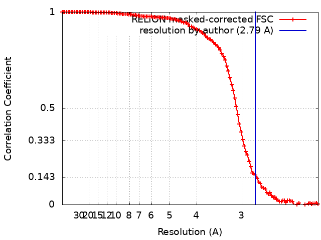
single particle reconstruction / cryo EM / Resolution: 2.79 Å
Name: POTASSIUM ION / type: ligand / ID: 75 / Number of copies: 199 / Formula: K
Molecular weight
Theoretical: 39.098 Da
+
Macromolecule #76: MAGNESIUM ION
Macromolecule
Name: MAGNESIUM ION / type: ligand / ID: 76 / Number of copies: 160 / Formula: MG
Molecular weight
Theoretical: 24.305 Da
+
Macromolecule #77: ZINC ION
Macromolecule
Name: ZINC ION / type: ligand / ID: 77 / Number of copies: 9 / Formula: ZN
Molecular weight
Theoretical: 65.409 Da
-
Experimental details
-
Structure determination
Method
cryo EM
Processing
single particle reconstruction
Aggregation state
particle
-
Sample preparation
Concentration
10 mg/mL
Buffer
pH: 7.5 Component:
Concentration
Formula
Name
25.0 mM
HEPES
HEPES
100.0 mM
CH3CO2K
Potassium Acetate
1.0 mM
DTT
Dithiothreitol
15.0 mM
Mg(C2H3O2)2
Magnessium Acetate
Details: Solution with HEPES, Potassium Acetate and Magnesium acetate was autoclaved. DTT was added fresh just before sample preparation.
Grid
Model: Quantifoil / Material: COPPER / Mesh: 300 / Support film - Material: CARBON / Support film - topology: CONTINUOUS / Support film - Film thickness: 2 / Pretreatment - Type: GLOW DISCHARGE / Pretreatment - Time: 1 sec. / Pretreatment - Atmosphere: OTHER / Pretreatment - Pressure: 0.026000000000000002 kPa / Details: 20 mA. Carbon coated grid
Vitrification
Cryogen name: ETHANE / Chamber humidity: 100 % / Chamber temperature: 288.15 K / Instrument: FEI VITROBOT MARK IV / Details: blot force -1 and blot time 4 s.
Details
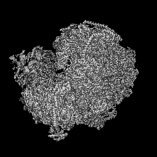
Monomeric ribosomes from Spraguea lophii
-
Electron microscopy #1
Microscopy ID
1
Microscope
TFS KRIOS
Image recording
Image recording ID: 1 / Film or detector model: GATAN K3 BIOQUANTUM (6k x 4k) / Average electron dose: 41.5 e/Å2
Electron beam
Acceleration voltage: 300 kV / Electron source: FIELD EMISSION GUN
In the structure databanks used in Yorodumi, some data are registered as the other names, "COVID-19 virus" and "2019-nCoV". Here are the details of the virus and the list of structure data.
Jan 31, 2019. EMDB accession codes are about to change! (news from PDBe EMDB page)
EMDB accession codes are about to change! (news from PDBe EMDB page)
The allocation of 4 digits for EMDB accession codes will soon come to an end. Whilst these codes will remain in use, new EMDB accession codes will include an additional digit and will expand incrementally as the available range of codes is exhausted. The current 4-digit format prefixed with “EMD-” (i.e. EMD-XXXX) will advance to a 5-digit format (i.e. EMD-XXXXX), and so on. It is currently estimated that the 4-digit codes will be depleted around Spring 2019, at which point the 5-digit format will come into force.
The EM Navigator/Yorodumi systems omit the EMD- prefix.
Related info.:Q: What is EMD? / ID/Accession-code notation in Yorodumi/EM Navigator
Yorodumi is a browser for structure data from EMDB, PDB, SASBDB, etc.
This page is also the successor to EM Navigator detail page, and also detail information page/front-end page for Omokage search.
The word "yorodu" (or yorozu) is an old Japanese word meaning "ten thousand". "mi" (miru) is to see.
Related info.:EMDB / PDB / SASBDB / Comparison of 3 databanks / Yorodumi Search / Aug 31, 2016. New EM Navigator & Yorodumi / Yorodumi Papers / Jmol/JSmol / Function and homology information / Changes in new EM Navigator and Yorodumi
 Movie
Movie Controller


 Open data
 Basic information
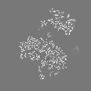
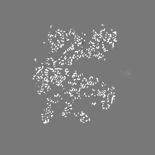
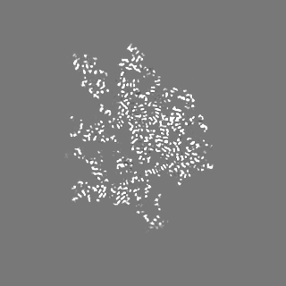
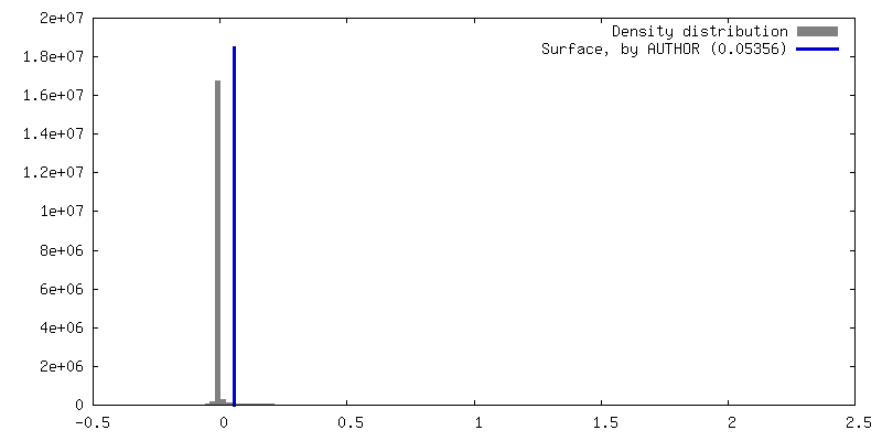
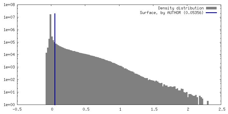
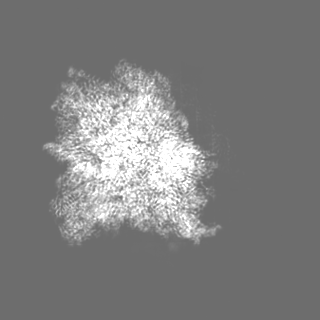
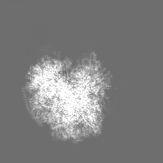
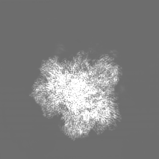
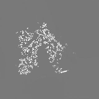
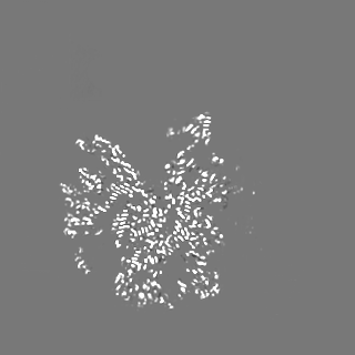
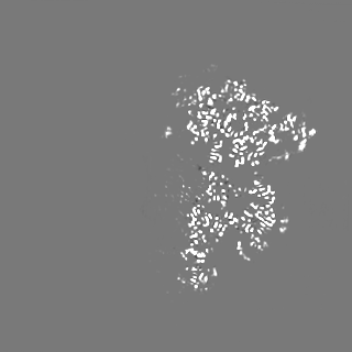
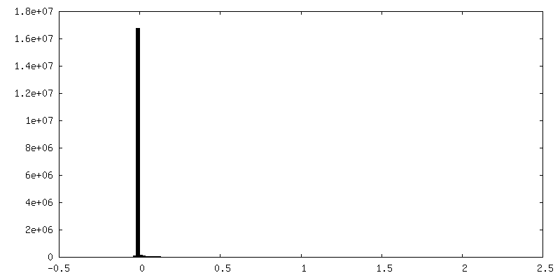
 Map data
 Sample
 Keywords
 Function and homology information
 Spraguea lophii 42_110 (fungus)
 Authors
 United Kingdom, 1 items
  Citation
 Structure visualization
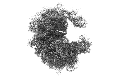
 Downloads & links
 emd_13892.png
 http://ftp.pdbj.org/pub/emdb/structures/EMD-13892


 Z (Sec.)
 Y (Row.)
 X (Col.)


 Sample components
 Processing
 Electron microscopy #1
 FIELD EMISSION GUN