 Movie
Movie Controller

[English] 日本語
 Yorodumi
- EMDB-1371: Architecture of the Dam1 kinetochore ring complex and implication... -
+ Open data

- Basic information
| Entry | Database: EMDB / ID: EMD-1371 | |||||||||
|---|---|---|---|---|---|---|---|---|---|---|
| Title | Architecture of the Dam1 kinetochore ring complex and implications for microtubule-driven assembly and force-coupling mechanisms. | |||||||||
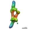
 Map data Map data | This is the reconstruction of Dam1 helical spiral around MT | |||||||||
 Sample Sample |
| |||||||||
| Biological species |  | |||||||||
| Method | helical reconstruction / cryo EM / Resolution: 30.0 Å | |||||||||
 Authors Authors | Wang H-W / Ramey VH / Westermann S / Leschziner AE / Welburn JPI / Nakajima Y / Drubin DG / Barnes G / Nogales E | |||||||||
 Citation Citation | Journal: Nat Struct Mol Biol / Year: 2007 Title: Architecture of the Dam1 kinetochore ring complex and implications for microtubule-driven assembly and force-coupling mechanisms. Authors: Hong-Wei Wang / Vincent H Ramey / Stefan Westermann / Andres E Leschziner / Julie P I Welburn / Yuko Nakajima / David G Drubin / Georjana Barnes / Eva Nogales /  Abstract: The Dam1 kinetochore complex is essential for chromosome segregation in budding yeast. This ten-protein complex self-assembles around microtubules, forming ring-like structures that move with ...The Dam1 kinetochore complex is essential for chromosome segregation in budding yeast. This ten-protein complex self-assembles around microtubules, forming ring-like structures that move with depolymerizing microtubule ends, a mechanism with implications for cellular function. Here we used EM-based single-particle and helical analyses to define the architecture of the Dam1 complex at 30-A resolution and the self-assembly mechanism. Ring oligomerization seems to be facilitated by a conformational change upon binding to microtubules, suggesting that the Dam1 ring is not preformed, but self-assembles around kinetochore microtubules. The C terminus of the Dam1p protein, where most of the Aurora kinase Ipl1 phosphorylation sites reside, is in a strategic location to affect oligomerization and interactions with the microtubule. One of Ipl1's roles might be to fine-tune the coupling of the microtubule interaction with the conformational change required for oligomerization, with phosphorylation resulting in ring breakdown. | |||||||||
| History |
|
- Structure visualization
| Movie |
 Movie viewer Movie viewer |
|---|---|
| Structure viewer | EM map: SurfViewMolmilJmol/JSmol |
| Supplemental images |
- Downloads & links
-EMDB archive
| Map data | emd_1371.map.gz | 3.2 MB | EMDB map data format | |
|---|---|---|---|---|
| Header (meta data) | emd-1371-v30.xmlemd-1371.xml | 11.1 KB 11.1 KB | Display Display | EMDB header |
| Images |  1371.gif 1371.gif | 100.3 KB | ||
| Archive directory |  http://ftp.pdbj.org/pub/emdb/structures/EMD-1371ftp://ftp.pdbj.org/pub/emdb/structures/EMD-1371 http://ftp.pdbj.org/pub/emdb/structures/EMD-1371ftp://ftp.pdbj.org/pub/emdb/structures/EMD-1371 | HTTPS FTP |
-Related structure data

-Links
| EMDB pages | EMDB (EBI/PDBe) / EMDataResource |
|---|
-Map
| File | Download / File: emd_1371.map.gz / Format: CCP4 / Size: 47.2 MB / Type: IMAGE STORED AS FLOATING POINT NUMBER (4 BYTES) | ||||||||||||||||||||||||||||||||||||||||||||||||||||||||||||
|---|---|---|---|---|---|---|---|---|---|---|---|---|---|---|---|---|---|---|---|---|---|---|---|---|---|---|---|---|---|---|---|---|---|---|---|---|---|---|---|---|---|---|---|---|---|---|---|---|---|---|---|---|---|---|---|---|---|---|---|---|---|
| Annotation | This is the reconstruction of Dam1 helical spiral around MT | ||||||||||||||||||||||||||||||||||||||||||||||||||||||||||||
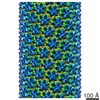
| Projections & slices | Image control
Images are generated by Spider. generated in cubic-lattice coordinate | ||||||||||||||||||||||||||||||||||||||||||||||||||||||||||||
| Voxel size | X=Y=Z: 4 Å | ||||||||||||||||||||||||||||||||||||||||||||||||||||||||||||
| Density |
| ||||||||||||||||||||||||||||||||||||||||||||||||||||||||||||
| Symmetry | Space group: 1 | ||||||||||||||||||||||||||||||||||||||||||||||||||||||||||||
| Details | EMDB XML:
CCP4 map header:
| ||||||||||||||||||||||||||||||||||||||||||||||||||||||||||||
 Z (Sec.)
 Y (Row.)
 X (Col.)


-Supplemental data
- Sample components
-Entire : Dam1 complex
| Entire | Name: Dam1 complex |
|---|---|
| Components |
|
-Supramolecule #1000: Dam1 complex
| Supramolecule | Name: Dam1 complex / type: sample / ID: 1000 Details: The well ordered long helical particles are rare while short helical assemblies with order are quite normal. Oligomeric state: Each assymmetric unit contains 10 subunits Number unique components: 1 |
|---|---|
| Molecular weight | Theoretical: 200 KDa |
-Macromolecule #1: Dam1 complex
| Macromolecule | Name: Dam1 complex / type: protein_or_peptide / ID: 1 / Name.synonym: DASH / Number of copies: 1 / Oligomeric state: decamer / Recombinant expression: Yes |
|---|---|
| Source (natural) | Organism: |
| Molecular weight | Experimental: 200 KDa / Theoretical: 200 KDa |
| Recombinant expression | Organism:  |
-Experimental details
-Structure determination
| Method | cryo EM |
|---|---|
 Processing Processing | helical reconstruction |
| Aggregation state | helical array |
-Sample preparation
| Concentration | 1 mg/mL |
|---|---|
| Buffer | pH: 6.8 Details: 150 mM KCl, 20 mM potassium phosphate pH 6.8, 1 mM EDTA |
| Grid | Details: 400 Quantifoil |
| Vitrification | Cryogen name: ETHANE / Chamber humidity: 100 % / Chamber temperature: 100 K / Instrument: OTHER Details: Vitrification instrument: Vitrobot. 3.5 micro-liter of sample solution was applied to glow-discharged Quantifoil and blotted with filter paper for 1.5 seconds and plunged Method: Blot for 1.5 seconds before plunging |
| Details | helical crystal grown in solution |
- Electron microscopy
| Microscope | FEI/PHILIPS CM200FEG |
|---|---|
| Temperature | Average: 100 K |
| Alignment procedure | Legacy - Astigmatism: objective lens astigmatism was corrected at |
| Details | Low Dose mode |
| Date | Nov 1, 2005 |
| Image recording | Category: FILM / Film or detector model: KODAK SO-163 FILM / Digitization - Scanner: OTHER / Digitization - Sampling interval: 12.7 µm / Number real images: 100 / Average electron dose: 15 e/Å2 / Details: Scanned on Nikon Super Coolscan 8000 / Od range: 1.5 / Bits/pixel: 14 |
| Electron beam | Acceleration voltage: 200 kV / Electron source:  FIELD EMISSION GUN FIELD EMISSION GUN |
| Electron optics | Illumination mode: FLOOD BEAM / Imaging mode: BRIGHT FIELD / Cs: 2.6 mm / Nominal defocus max: 2.0 µm / Nominal defocus min: 1.5 µm / Nominal magnification: 50000 |
| Sample stage | Specimen holder: Side-entry liquid nitrogen cooled cryo-holder Specimen holder model: GATAN LIQUID NITROGEN |
-Image processing
| Details | helices were formed around microtubules |
|---|---|
| Final reconstruction | Applied symmetry - Helical parameters - Δz: 10.2 Å Applied symmetry - Helical parameters - Δ&Phi: 24.7 ° Applied symmetry - Helical parameters - Axial symmetry: D2 (2x2 fold dihedral) Algorithm: OTHER / Resolution.type: BY AUTHOR / Resolution: 30.0 Å / Resolution method: OTHER / Software - Name: SUPRIM, MRC Details: Final map was calculated from two particle images. The two images were of different helical families, thus the average in little g space was performed. |
