 Movie
Movie Controller


+ Open data

- Basic information
| Entry | 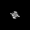 | |||||||||
|---|---|---|---|---|---|---|---|---|---|---|
| Title | CryoEM structure of DnaD dimer from Bacillus subtilis | |||||||||
 Map data Map data | Structure of the dimeric state of DnaD | |||||||||
 Sample Sample |
| |||||||||
 Keywords Keywords | Replication / Initiation / Bacteria / Bacillus subtilis / DNA BINDING PROTEIN | |||||||||
| Biological species |  | |||||||||
| Method | single particle reconstruction / cryo EM / Resolution: 10.1 Å | |||||||||
 Authors Authors | Winterhalter C / Pelliciari S / Cronin N / Costa TRD / Ilangovan I / Murray H | |||||||||
| Funding support |  United Kingdom, 1 items United Kingdom, 1 items
| |||||||||
 Citation Citation | Journal: Biorxiv / Year: 2022 Title: The DNA replication initiation protein DnaD is recruited to a specific strand of the Bacillus subtilis chromosome origin Authors: Winterhalter C / Pelliciari S / Stevens D / Fenyk S / Marchand E / Cronin NB / Soultanas P / Costa TRD / Ilangovan A / Murray H | |||||||||
| History |
|
- Structure visualization
| Supplemental images |
|---|

- Downloads & links
-EMDB archive
| Map data | emd_13663.map.gz | 86.6 MB |  EMDB map data format EMDB map data format | |
|---|---|---|---|---|
| Header (meta data) | emd-13663-v30.xmlemd-13663.xml | 7.8 KB 7.8 KB | Display Display | EMDB header |
| Images |  emd_13663.png emd_13663.png | 25.4 KB | ||
| Filedesc metadata | emd-13663.cif.gz | 3.6 KB | ||
| Archive directory |  http://ftp.pdbj.org/pub/emdb/structures/EMD-13663ftp://ftp.pdbj.org/pub/emdb/structures/EMD-13663 http://ftp.pdbj.org/pub/emdb/structures/EMD-13663ftp://ftp.pdbj.org/pub/emdb/structures/EMD-13663 | HTTPS FTP |
-Links
| EMDB pages | EMDB (EBI/PDBe) / EMDataResource |
|---|
-Map
| File | Download / File: emd_13663.map.gz / Format: CCP4 / Size: 178 MB / Type: IMAGE STORED AS FLOATING POINT NUMBER (4 BYTES) | ||||||||||||||||||||||||||||||||||||
|---|---|---|---|---|---|---|---|---|---|---|---|---|---|---|---|---|---|---|---|---|---|---|---|---|---|---|---|---|---|---|---|---|---|---|---|---|---|
| Annotation | Structure of the dimeric state of DnaD | ||||||||||||||||||||||||||||||||||||
| Projections & slices | Image control
Images are generated by Spider. | ||||||||||||||||||||||||||||||||||||
| Voxel size | X=Y=Z: 0.67 Å | ||||||||||||||||||||||||||||||||||||
| Density |
| ||||||||||||||||||||||||||||||||||||
| Symmetry | Space group: 1 | ||||||||||||||||||||||||||||||||||||
| Details | EMDB XML:
|
 Z (Sec.)
 Y (Row.)
 X (Col.)


-Supplemental data
- Sample components
-Entire : Dimer of the replication initiation protein DnaD
| Entire | Name: Dimer of the replication initiation protein DnaD |
|---|---|
| Components |
|
-Supramolecule #1: Dimer of the replication initiation protein DnaD
| Supramolecule | Name: Dimer of the replication initiation protein DnaD / type: complex / ID: 1 / Parent: 0 |
|---|---|
| Source (natural) | Organism: |
| Molecular weight | Theoretical: 54 KDa |
-Experimental details
-Structure determination
| Method | cryo EM |
|---|---|
 Processing Processing | single particle reconstruction |
| Aggregation state | particle |
-Sample preparation
| Buffer | pH: 7.5 |
|---|---|
| Vitrification | Cryogen name: ETHANE / Instrument: LEICA EM GP |
- Electron microscopy
| Microscope | FEI TITAN KRIOS |
|---|---|
| Image recording | Film or detector model: GATAN K3 (6k x 4k) / Average electron dose: 50.0 e/Å2 |
| Electron beam | Acceleration voltage: 300 kV / Electron source:  FIELD EMISSION GUN FIELD EMISSION GUN |
| Electron optics | Illumination mode: SPOT SCAN / Imaging mode: BRIGHT FIELD |
| Experimental equipment |  Model: Titan Krios / Image courtesy: FEI Company |
-Image processing
| Startup model | Type of model: OTHER |
|---|---|
| Final reconstruction | Resolution.type: BY AUTHOR / Resolution: 10.1 Å / Resolution method: FSC 0.5 CUT-OFF / Number images used: 73190 |
| Initial angle assignment | Type: MAXIMUM LIKELIHOOD |
| Final angle assignment | Type: MAXIMUM LIKELIHOOD |
