 Movie
Movie Controller

[English] 日本語
 Yorodumi
- EMDB-13585: Cryo-electron tomography of ASC signalling sites in pyroptotic cells -
+ Open data

- Basic information
| Entry |  | |||||||||
|---|---|---|---|---|---|---|---|---|---|---|
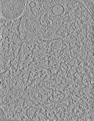
| Title | Cryo-electron tomography of ASC signalling sites in pyroptotic cells | |||||||||
 Map data Map data | ||||||||||
 Sample Sample |
| |||||||||
| Biological species |  | |||||||||
| Method | electron tomography / cryo EM | |||||||||
 Authors Authors | Liu Y / Zhai H / Alemayehu H / Hopkins LJ / Borgeaud AC / Heroven C / Howe JD / Boulanger J / Bryant CE / Modis Y | |||||||||
| Funding support |  United Kingdom, 1 items United Kingdom, 1 items
| |||||||||
 Citation Citation | Journal: To Be Published Title: Cryo-electron tomography of ASC signalling sites in pyroptotic cells Authors: Liu Y / Zhai H / Alemayehu H / Hopkins LJ / Borgeaud AC / Heroven C / Howe JD / Boulanger J / Bryant CE / Modis Y | |||||||||
| History |
|
- Structure visualization
| Supplemental images |
|---|
- Downloads & links
-EMDB archive
| Map data | emd_13585.map.gz | 704.8 MB |  EMDB map data format EMDB map data format | |
|---|---|---|---|---|
| Header (meta data) | emd-13585-v30.xmlemd-13585.xml | 9.4 KB 9.4 KB | Display Display | EMDB header |
| Images |  emd_13585.png emd_13585.png | 190.7 KB | ||
| Archive directory |  http://ftp.pdbj.org/pub/emdb/structures/EMD-13585ftp://ftp.pdbj.org/pub/emdb/structures/EMD-13585 http://ftp.pdbj.org/pub/emdb/structures/EMD-13585ftp://ftp.pdbj.org/pub/emdb/structures/EMD-13585 | HTTPS FTP |
-Validation report
| Summary document | emd_13585_validation.pdf.gz | 397.9 KB | Display | EMDB validaton report |
|---|---|---|---|---|
| Full document | emd_13585_full_validation.pdf.gz | 397.4 KB | Display | |
| Data in XML | emd_13585_validation.xml.gz | 4.8 KB | Display | |
| Data in CIF | emd_13585_validation.cif.gz | 5.3 KB | Display | |
| Arichive directory | https://ftp.pdbj.org/pub/emdb/validation_reports/EMD-13585ftp://ftp.pdbj.org/pub/emdb/validation_reports/EMD-13585 | HTTPS FTP |
-Links
| EMDB pages | EMDB (EBI/PDBe) / EMDataResource |
|---|
-Map
| File | Download / File: emd_13585.map.gz / Format: CCP4 / Size: 760.2 MB / Type: IMAGE STORED AS FLOATING POINT NUMBER (4 BYTES) | ||||||||||||||||||||||||||||||||
|---|---|---|---|---|---|---|---|---|---|---|---|---|---|---|---|---|---|---|---|---|---|---|---|---|---|---|---|---|---|---|---|---|---|
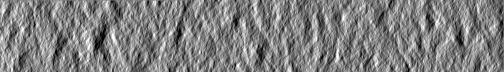
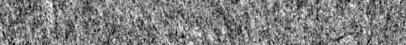
| Projections & slices | Image control
Images are generated by Spider. generated in cubic-lattice coordinate | ||||||||||||||||||||||||||||||||
| Voxel size | X=Y=Z: 13.58 Å | ||||||||||||||||||||||||||||||||
| Density |
| ||||||||||||||||||||||||||||||||
| Symmetry | Space group: 1 | ||||||||||||||||||||||||||||||||
| Details | EMDB XML:
|
 Z (Sec.)
 Y (Row.)
 X (Col.)


-Supplemental data
- Sample components
-Entire : FIB milled iBMDMs
| Entire | Name: FIB milled iBMDMs |
|---|---|
| Components |
|
-Supramolecule #1: FIB milled iBMDMs
| Supramolecule | Name: FIB milled iBMDMs / type: cell / ID: 1 / Parent: 0 |
|---|---|
| Source (natural) | Organism: |
-Experimental details
-Structure determination
| Method | cryo EM |
|---|---|
 Processing Processing | electron tomography |
| Aggregation state | cell |
-Sample preparation
| Buffer | pH: 7.4 Details: DMEM medium supplemented with 10% heat-inactivated FBS (Gibco) |
|---|---|
| Grid | Model: Quantifoil R2/1 / Material: GOLD / Mesh: 200 / Support film - Material: CARBON / Support film - topology: HOLEY / Pretreatment - Type: GLOW DISCHARGE / Pretreatment - Atmosphere: AIR |
| Vitrification | Cryogen name: ETHANE / Chamber humidity: 100 % / Chamber temperature: 298 K / Instrument: LEICA EM GP |
| Sectioning | Focused ion beam - Instrument: OTHER / Focused ion beam - Ion: OTHER / Focused ion beam - Voltage: 30 kV / Focused ion beam - Current: 1 nA / Focused ion beam - Duration: 1500 sec. / Focused ion beam - Temperature: 83 K / Focused ion beam - Initial thickness: 10000 nm / Focused ion beam - Final thickness: 200 nm Focused ion beam - Details: Lamella were made using a Scios DualBeam FIB/SEM (FEI) equipped with a Quorum cryo-stage (PP3010T). Rough milling was performed using 0.5 to 0.1 nA current. Fine milling ...Focused ion beam - Details: Lamella were made using a Scios DualBeam FIB/SEM (FEI) equipped with a Quorum cryo-stage (PP3010T). Rough milling was performed using 0.5 to 0.1 nA current. Fine milling was done using 50 to 10 pA current.. |
- Electron microscopy
| Microscope | FEI TITAN KRIOS |
|---|---|
| Image recording | Film or detector model: GATAN K3 (6k x 4k) / Number real images: 121 / Average electron dose: 1.3 e/Å2 |
| Electron beam | Acceleration voltage: 300 kV / Electron source:  FIELD EMISSION GUN FIELD EMISSION GUN |
| Electron optics | Illumination mode: FLOOD BEAM / Imaging mode: BRIGHT FIELD / Nominal defocus max: 7.0 µm / Nominal defocus min: 4.0 µm |
| Sample stage | Specimen holder model: FEI TITAN KRIOS AUTOGRID HOLDER / Cooling holder cryogen: NITROGEN |
| Experimental equipment |  Model: Titan Krios / Image courtesy: FEI Company |
-Image processing
| Final reconstruction | Resolution method: OTHER / Software - Name: eTomo / Number images used: 100 |
|---|---|
| CTF correction | Software - Name: eTomo (ver. 4.10.20) |
