 ムービー
ムービー コントローラー


+ データを開く

- 基本情報
基本情報
| 登録情報 | データベース: EMDB / ID: EMD-1355 | |||||||||
|---|---|---|---|---|---|---|---|---|---|---|
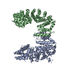
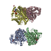
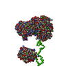
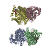
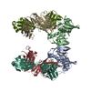
| タイトル | Molecular architecture of the human GINS complex. | |||||||||
 マップデータ マップデータ | This is an image of a surface of the human GINS complex | |||||||||
 試料 試料 |
| |||||||||
| 機能・相同性 | DNA replication complex GINS protein Psf2 / GINS complex, subunit Psf3 / GINS complex subunit Sld5 / GINS complex, subunit Psf1 機能・相同性情報 機能・相同性情報 | |||||||||
| 生物種 |  Homo sapiens (ヒト) Homo sapiens (ヒト) | |||||||||
| 手法 | 単粒子再構成法 / ネガティブ染色法 / 解像度: 33.0 Å | |||||||||
 データ登録者 データ登録者 | Boskovic J / Coloma J / Aparicio T / Zhou M / Robinson CV / Mendez J / Montoya G | |||||||||
 引用 引用 | ジャーナル: EMBO Rep / 年: 2007 タイトル: Molecular architecture of the human GINS complex. 著者: Jasminka Boskovic / Javier Coloma / Tomás Aparicio / Min Zhou / Carol V Robinson / Juan Méndez / Guillermo Montoya /  要旨: Chromosomal DNA replication is strictly regulated through a sequence of steps that involve many macromolecular protein complexes. One of these is the GINS complex, which is required for initiation ...Chromosomal DNA replication is strictly regulated through a sequence of steps that involve many macromolecular protein complexes. One of these is the GINS complex, which is required for initiation and elongation phases in eukaryotic DNA replication. The GINS complex consists of four paralogous subunits. At the G1/S transition, GINS is recruited to the origins of replication where it assembles with cell-division cycle protein (Cdc)45 and the minichromosome maintenance mutant (MCM)2-7 to form the Cdc45/Mcm2-7/GINS (CMG) complex, the presumed replicative helicase. We isolated the human GINS complex and have shown that it can bind to DNA. By using single-particle electron microscopy and three-dimensional reconstruction, we obtained a medium-resolution volume of the human GINS complex, which shows a horseshoe shape. Analysis of the protein interactions using mass spectrometry and monoclonal antibody mapping shows the subunit organization within the GINS complex. The structure and DNA-binding data suggest how GINS could interact with DNA and also its possible role in the CMG helicase complex. | |||||||||
| 履歴 |
|
- 構造の表示
構造の表示
| ムービー |
ムービービューア |
|---|---|
| 構造ビューア | EMマップ: SurfViewMolmilJmol/JSmol |
| 添付画像 |
 UCSF Chimera
- ダウンロードとリンク
-EMDBアーカイブ
| マップデータ | emd_1355.map.gz | 4.6 MB | EMDBマップデータ形式 | |
|---|---|---|---|---|
| ヘッダ (付随情報) | emd-1355-v30.xmlemd-1355.xml | 12.4 KB 12.4 KB | 表示 表示 | EMDBヘッダ |
| 画像 |  1355.gif 1355.gif | 56.2 KB | ||
| アーカイブディレクトリ |  http://ftp.pdbj.org/pub/emdb/structures/EMD-1355ftp://ftp.pdbj.org/pub/emdb/structures/EMD-1355 http://ftp.pdbj.org/pub/emdb/structures/EMD-1355ftp://ftp.pdbj.org/pub/emdb/structures/EMD-1355 | HTTPS FTP |
-検証レポート
| 文書・要旨 | emd_1355_validation.pdf.gz | 197 KB | 表示 | EMDB検証レポート |
|---|---|---|---|---|
| 文書・詳細版 | emd_1355_full_validation.pdf.gz | 196.1 KB | 表示 | |
| XML形式データ | emd_1355_validation.xml.gz | 5.6 KB | 表示 | |
| アーカイブディレクトリ | https://ftp.pdbj.org/pub/emdb/validation_reports/EMD-1355ftp://ftp.pdbj.org/pub/emdb/validation_reports/EMD-1355 | HTTPS FTP |
-関連構造データ

-リンク
| EMDBのページ | EMDB (EBI/PDBe) / EMDataResource |
|---|
-マップ
| ファイル | ダウンロード / ファイル: emd_1355.map.gz / 形式: CCP4 / 大きさ: 5.2 MB / タイプ: IMAGE STORED AS FLOATING POINT NUMBER (4 BYTES) | ||||||||||||||||||||||||||||||||||||||||||||||||||||||||||||||||||||
|---|---|---|---|---|---|---|---|---|---|---|---|---|---|---|---|---|---|---|---|---|---|---|---|---|---|---|---|---|---|---|---|---|---|---|---|---|---|---|---|---|---|---|---|---|---|---|---|---|---|---|---|---|---|---|---|---|---|---|---|---|---|---|---|---|---|---|---|---|---|
| 注釈 | This is an image of a surface of the human GINS complex | ||||||||||||||||||||||||||||||||||||||||||||||||||||||||||||||||||||
| 投影像・断面図 | 画像のコントロール
画像は Spider により作成 | ||||||||||||||||||||||||||||||||||||||||||||||||||||||||||||||||||||
| ボクセルのサイズ | X=Y=Z: 3.56 Å | ||||||||||||||||||||||||||||||||||||||||||||||||||||||||||||||||||||
| 密度 |
| ||||||||||||||||||||||||||||||||||||||||||||||||||||||||||||||||||||
| 対称性 | 空間群: 1 | ||||||||||||||||||||||||||||||||||||||||||||||||||||||||||||||||||||
| 詳細 | EMDB XML:
CCP4マップ ヘッダ情報:
| ||||||||||||||||||||||||||||||||||||||||||||||||||||||||||||||||||||
 Z (Sec.)
 Y (Row.)
 X (Col.)


-添付データ
- 試料の構成要素
-全体 : Human GINS complex
| 全体 | 名称: Human GINS complex |
|---|---|
| 要素 |
|
-超分子 #1000: Human GINS complex
| 超分子 | 名称: Human GINS complex / タイプ: sample / ID: 1000 / 詳細: The sample was monodisperse / 集合状態: Monomer / Number unique components: 4 |
|---|---|
| 分子量 | 実験値: 983.73 KDa / 理論値: 981.22 KDa / 手法: Nano-flow MS of the intact complex |
-分子 #1: hPsf1p
| 分子 | 名称: hPsf1p / タイプ: protein_or_peptide / ID: 1 / Name.synonym: GINS1 / コピー数: 1 / 集合状態: Monomer / 組換発現: Yes |
|---|---|
| 由来(天然) | 生物種: Homo sapiens (ヒト) / 別称: Human / Organelle: Nucleus |
| 分子量 | 実験値: 229.88 KDa |
| 組換発現 | 生物種:  |
| 配列 | InterPro: GINS complex, subunit Psf1 |
-分子 #2: hPsf2
| 分子 | 名称: hPsf2 / タイプ: protein_or_peptide / ID: 2 / Name.synonym: GINS2 / コピー数: 1 / 集合状態: Monomer / 組換発現: Yes |
|---|---|
| 由来(天然) | 生物種: Homo sapiens (ヒト) / 別称: Human / Organelle: Nucleus |
| 分子量 | 実験値: 214.28 KDa |
| 組換発現 | 生物種: |
| 配列 | InterPro: DNA replication complex GINS protein Psf2 |
-分子 #3: hPsf3
| 分子 | 名称: hPsf3 / タイプ: protein_or_peptide / ID: 3 / Name.synonym: GINS3 / 詳細: Hexa His-tag on its N-terminal / コピー数: 1 / 集合状態: Monomer / 組換発現: Yes |
|---|---|
| 由来(天然) | 生物種: Homo sapiens (ヒト) / 別称: Human / Organelle: Nucleus |
| 分子量 | 実験値: 240 KDa |
| 組換発現 | 生物種: |
| 配列 | InterPro: GINS complex, subunit Psf3 |
-分子 #4: hSld5
| 分子 | 名称: hSld5 / タイプ: protein_or_peptide / ID: 4 / Name.synonym: GINS4 / コピー数: 1 / 集合状態: Monomer / 組換発現: Yes |
|---|---|
| 由来(天然) | 生物種: Homo sapiens (ヒト) / 別称: Human / Organelle: Nucleus |
| 分子量 | 実験値: 260.47 KDa |
| 組換発現 | 生物種: |
| 配列 | InterPro: GINS complex subunit Sld5 |
-実験情報
-構造解析
| 手法 | ネガティブ染色法 |
|---|---|
 解析 解析 | 単粒子再構成法 |
| 試料の集合状態 | particle |
-試料調製
| 濃度 | 0.1 mg/mL |
|---|---|
| 緩衝液 | pH: 8 詳細: 10 mM Tris-HCl pH 8.0, 100 mM NaCl, 0.5 mM EDTA, 10% glycerol, 1 mM DTT |
| 染色 | タイプ: NEGATIVE 詳細: Grids with adsorbed protein were stained with 0.2 % uranyl acetate for 1 min |
| グリッド | 詳細: 400 mesh copper-rhodium grid |
| 凍結 | 凍結剤: NONE |
- 電子顕微鏡法
| 顕微鏡 | JEOL 1230 |
|---|---|
| アライメント法 | Legacy - 非点収差: objective lens astigmatism was corrected at 100,000 times magnification |
| 詳細 | Electron microscope was JEOL JEM-120, 120kV. Specimen holder, JEOL type M: 207EM-11020. Images were colected in low-dose conditions |
| 日付 | 2007年11月18日 |
| 撮影 | カテゴリ: FILM / フィルム・検出器のモデル: KODAK 4489 FILM / デジタル化 - スキャナー: OTHER / デジタル化 - サンプリング間隔: 3.56 µm / 実像数: 40 / 詳細: Minolta Dimage Scan Multi Pro. Scan at 2400 dpi / ビット/ピクセル: 16 |
| Tilt angle min | 0 |
| 電子線 | 加速電圧: 100 kV / 電子線源: TUNGSTEN HAIRPIN |
| 電子光学系 | 照射モード: SPOT SCAN / 撮影モード: DIFFRACTION / Cs: 2.9 mm / 倍率(公称値): 60000 |
| 試料ステージ | 試料ホルダー: Eucentric / 試料ホルダーモデル: OTHER / Tilt angle max: 20 |
-画像解析
| CTF補正 | 詳細: Each micrograph |
|---|---|
| 最終 再構成 | 想定した対称性 - 点群: C1 (非対称) / アルゴリズム: OTHER / 解像度のタイプ: BY AUTHOR / 解像度: 33.0 Å / 解像度の算出法: FSC 0.5 CUT-OFF / ソフトウェア - 名称: EMAN / 使用した粒子像数: 5290 |
