 Movie
Movie Controller


+ Open data

- Basic information
| Entry | Database: EMDB / ID: EMD-1355 | |||||||||
|---|---|---|---|---|---|---|---|---|---|---|
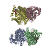
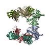
| Title | Molecular architecture of the human GINS complex. | |||||||||
 Map data Map data | This is an image of a surface of the human GINS complex | |||||||||
 Sample Sample |
| |||||||||
| Function / homology | DNA replication complex GINS protein Psf2 / GINS complex, subunit Psf3 / GINS complex subunit Sld5 / GINS complex, subunit Psf1 Function and homology information Function and homology information | |||||||||
| Biological species |  Homo sapiens (human) Homo sapiens (human) | |||||||||
| Method | single particle reconstruction / negative staining / Resolution: 33.0 Å | |||||||||
 Authors Authors | Boskovic J / Coloma J / Aparicio T / Zhou M / Robinson CV / Mendez J / Montoya G | |||||||||
 Citation Citation | Journal: EMBO Rep / Year: 2007 Title: Molecular architecture of the human GINS complex. Authors: Jasminka Boskovic / Javier Coloma / Tomás Aparicio / Min Zhou / Carol V Robinson / Juan Méndez / Guillermo Montoya /  Abstract: Chromosomal DNA replication is strictly regulated through a sequence of steps that involve many macromolecular protein complexes. One of these is the GINS complex, which is required for initiation ...Chromosomal DNA replication is strictly regulated through a sequence of steps that involve many macromolecular protein complexes. One of these is the GINS complex, which is required for initiation and elongation phases in eukaryotic DNA replication. The GINS complex consists of four paralogous subunits. At the G1/S transition, GINS is recruited to the origins of replication where it assembles with cell-division cycle protein (Cdc)45 and the minichromosome maintenance mutant (MCM)2-7 to form the Cdc45/Mcm2-7/GINS (CMG) complex, the presumed replicative helicase. We isolated the human GINS complex and have shown that it can bind to DNA. By using single-particle electron microscopy and three-dimensional reconstruction, we obtained a medium-resolution volume of the human GINS complex, which shows a horseshoe shape. Analysis of the protein interactions using mass spectrometry and monoclonal antibody mapping shows the subunit organization within the GINS complex. The structure and DNA-binding data suggest how GINS could interact with DNA and also its possible role in the CMG helicase complex. | |||||||||
| History |
|
- Structure visualization
| Movie |
Movie viewer |
|---|---|
| Structure viewer | EM map: SurfViewMolmilJmol/JSmol |
| Supplemental images |
 UCSF Chimera
- Downloads & links
-EMDB archive
| Map data | emd_1355.map.gz | 4.6 MB | EMDB map data format | |
|---|---|---|---|---|
| Header (meta data) | emd-1355-v30.xmlemd-1355.xml | 12.4 KB 12.4 KB | Display Display | EMDB header |
| Images | 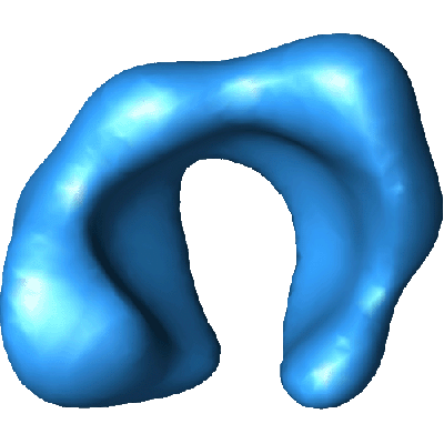 1355.gif 1355.gif | 56.2 KB | ||
| Archive directory |  http://ftp.pdbj.org/pub/emdb/structures/EMD-1355ftp://ftp.pdbj.org/pub/emdb/structures/EMD-1355 http://ftp.pdbj.org/pub/emdb/structures/EMD-1355ftp://ftp.pdbj.org/pub/emdb/structures/EMD-1355 | HTTPS FTP |
-Related structure data
| Similar structure data |
|---|

-Links
| EMDB pages | EMDB (EBI/PDBe) / EMDataResource |
|---|
-Map
| File | Download / File: emd_1355.map.gz / Format: CCP4 / Size: 5.2 MB / Type: IMAGE STORED AS FLOATING POINT NUMBER (4 BYTES) | ||||||||||||||||||||||||||||||||||||||||||||||||||||||||||||||||||||
|---|---|---|---|---|---|---|---|---|---|---|---|---|---|---|---|---|---|---|---|---|---|---|---|---|---|---|---|---|---|---|---|---|---|---|---|---|---|---|---|---|---|---|---|---|---|---|---|---|---|---|---|---|---|---|---|---|---|---|---|---|---|---|---|---|---|---|---|---|---|
| Annotation | This is an image of a surface of the human GINS complex | ||||||||||||||||||||||||||||||||||||||||||||||||||||||||||||||||||||
| Projections & slices | Image control
Images are generated by Spider. | ||||||||||||||||||||||||||||||||||||||||||||||||||||||||||||||||||||
| Voxel size | X=Y=Z: 3.56 Å | ||||||||||||||||||||||||||||||||||||||||||||||||||||||||||||||||||||
| Density |
| ||||||||||||||||||||||||||||||||||||||||||||||||||||||||||||||||||||
| Symmetry | Space group: 1 | ||||||||||||||||||||||||||||||||||||||||||||||||||||||||||||||||||||
| Details | EMDB XML:
CCP4 map header:
| ||||||||||||||||||||||||||||||||||||||||||||||||||||||||||||||||||||
 Z (Sec.)
 Y (Row.)
 X (Col.)


-Supplemental data
- Sample components
-Entire : Human GINS complex
| Entire | Name: Human GINS complex |
|---|---|
| Components |
|
-Supramolecule #1000: Human GINS complex
| Supramolecule | Name: Human GINS complex / type: sample / ID: 1000 / Details: The sample was monodisperse / Oligomeric state: Monomer / Number unique components: 4 |
|---|---|
| Molecular weight | Experimental: 983.73 KDa / Theoretical: 981.22 KDa / Method: Nano-flow MS of the intact complex |
-Macromolecule #1: hPsf1p
| Macromolecule | Name: hPsf1p / type: protein_or_peptide / ID: 1 / Name.synonym: GINS1 / Number of copies: 1 / Oligomeric state: Monomer / Recombinant expression: Yes |
|---|---|
| Source (natural) | Organism: Homo sapiens (human) / synonym: Human / Organelle: Nucleus |
| Molecular weight | Experimental: 229.88 KDa |
| Recombinant expression | Organism:  |
| Sequence | InterPro: GINS complex, subunit Psf1 |
-Macromolecule #2: hPsf2
| Macromolecule | Name: hPsf2 / type: protein_or_peptide / ID: 2 / Name.synonym: GINS2 / Number of copies: 1 / Oligomeric state: Monomer / Recombinant expression: Yes |
|---|---|
| Source (natural) | Organism: Homo sapiens (human) / synonym: Human / Organelle: Nucleus |
| Molecular weight | Experimental: 214.28 KDa |
| Recombinant expression | Organism: |
| Sequence | InterPro: DNA replication complex GINS protein Psf2 |
-Macromolecule #3: hPsf3
| Macromolecule | Name: hPsf3 / type: protein_or_peptide / ID: 3 / Name.synonym: GINS3 / Details: Hexa His-tag on its N-terminal / Number of copies: 1 / Oligomeric state: Monomer / Recombinant expression: Yes |
|---|---|
| Source (natural) | Organism: Homo sapiens (human) / synonym: Human / Organelle: Nucleus |
| Molecular weight | Experimental: 240 KDa |
| Recombinant expression | Organism: |
| Sequence | InterPro: GINS complex, subunit Psf3 |
-Macromolecule #4: hSld5
| Macromolecule | Name: hSld5 / type: protein_or_peptide / ID: 4 / Name.synonym: GINS4 / Number of copies: 1 / Oligomeric state: Monomer / Recombinant expression: Yes |
|---|---|
| Source (natural) | Organism: Homo sapiens (human) / synonym: Human / Organelle: Nucleus |
| Molecular weight | Experimental: 260.47 KDa |
| Recombinant expression | Organism: |
| Sequence | InterPro: GINS complex subunit Sld5 |
-Experimental details
-Structure determination
| Method | negative staining |
|---|---|
 Processing Processing | single particle reconstruction |
| Aggregation state | particle |
-Sample preparation
| Concentration | 0.1 mg/mL |
|---|---|
| Buffer | pH: 8 Details: 10 mM Tris-HCl pH 8.0, 100 mM NaCl, 0.5 mM EDTA, 10% glycerol, 1 mM DTT |
| Staining | Type: NEGATIVE Details: Grids with adsorbed protein were stained with 0.2 % uranyl acetate for 1 min |
| Grid | Details: 400 mesh copper-rhodium grid |
| Vitrification | Cryogen name: NONE |
- Electron microscopy
| Microscope | JEOL 1230 |
|---|---|
| Alignment procedure | Legacy - Astigmatism: objective lens astigmatism was corrected at 100,000 times magnification |
| Details | Electron microscope was JEOL JEM-120, 120kV. Specimen holder, JEOL type M: 207EM-11020. Images were colected in low-dose conditions |
| Date | Nov 18, 2007 |
| Image recording | Category: FILM / Film or detector model: KODAK 4489 FILM / Digitization - Scanner: OTHER / Digitization - Sampling interval: 3.56 µm / Number real images: 40 / Details: Minolta Dimage Scan Multi Pro. Scan at 2400 dpi / Bits/pixel: 16 |
| Tilt angle min | 0 |
| Electron beam | Acceleration voltage: 100 kV / Electron source: TUNGSTEN HAIRPIN |
| Electron optics | Illumination mode: SPOT SCAN / Imaging mode: DIFFRACTION / Cs: 2.9 mm / Nominal magnification: 60000 |
| Sample stage | Specimen holder: Eucentric / Specimen holder model: OTHER / Tilt angle max: 20 |
-Image processing
| CTF correction | Details: Each micrograph |
|---|---|
| Final reconstruction | Applied symmetry - Point group: C1 (asymmetric) / Algorithm: OTHER / Resolution.type: BY AUTHOR / Resolution: 33.0 Å / Resolution method: FSC 0.5 CUT-OFF / Software - Name: EMAN / Number images used: 5290 |
