 Movie
Movie Controller

[English] 日本語
 Yorodumi
- EMDB-1354: Allosteric signaling and a nuclear exit strategy: binding of UL25... -
+ Open data

- Basic information
| Entry | Database: EMDB / ID: EMD-1354 | |||||||||
|---|---|---|---|---|---|---|---|---|---|---|
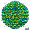
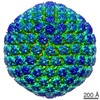
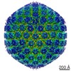
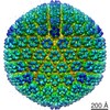
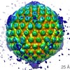
| Title | Allosteric signaling and a nuclear exit strategy: binding of UL25/UL17 heterodimers to DNA-Filled HSV-1 capsids. | |||||||||
 Map data Map data | EM Map from HSV-1 C-capsids | |||||||||
 Sample Sample |
| |||||||||
| Biological species |   Human herpesvirus 1 (Herpes simplex virus type 1) Human herpesvirus 1 (Herpes simplex virus type 1) | |||||||||
| Method | single particle reconstruction / cryo EM / Resolution: 19.9 Å | |||||||||
 Authors Authors | Trus BL / Newcomb WW / Cheng N / Cardone G / Marekov L / Homa FL / Brown JC / Steven AC | |||||||||
 Citation Citation | Journal: Mol Cell / Year: 2007 Title: Allosteric signaling and a nuclear exit strategy: binding of UL25/UL17 heterodimers to DNA-Filled HSV-1 capsids. Authors: Benes L Trus / William W Newcomb / Naiqian Cheng / Giovanni Cardone / Lyuben Marekov / Fred L Homa / Jay C Brown / Alasdair C Steven /  Abstract: UL25 and UL17 are two essential minor capsid proteins of HSV-1, implicated in DNA packaging and capsid maturation. We used cryo-electron microscopy to examine their binding to capsids, whose ...UL25 and UL17 are two essential minor capsid proteins of HSV-1, implicated in DNA packaging and capsid maturation. We used cryo-electron microscopy to examine their binding to capsids, whose architecture observes T = 16 icosahedral geometry. C-capsids (mature DNA-filled capsids) have an elongated two-domain molecule present at a unique, vertex-adjacent site that is not seen at other quasiequivalent sites or on unfilled capsids. Using SDS-PAGE and mass spectrometry to analyze wild-type capsids, UL25 null capsids, and denaturant-extracted capsids, we conclude that (1) the C-capsid-specific component is a heterodimer of UL25 and UL17, and (2) capsids have additional populations of UL25 and UL17 that are invisible in reconstructions because of sparsity and/or disorder. We infer that binding of the ordered population reflects structural changes induced on the outer surface as pressure builds up inside the capsid during DNA packaging. Its binding may signal that the C-capsid is ready to exit the nucleus. | |||||||||
| History |
|
- Structure visualization
| Movie |
 Movie viewer Movie viewer |
|---|---|
| Structure viewer | EM map: SurfViewMolmilJmol/JSmol |
| Supplemental images |
- Downloads & links
-EMDB archive
| Map data | emd_1354.map.gz | 113.3 MB | EMDB map data format | |
|---|---|---|---|---|
| Header (meta data) | emd-1354-v30.xmlemd-1354.xml | 9.4 KB 9.4 KB | Display Display | EMDB header |
| Images |  1354.gif 1354.gif | 26.1 KB | ||
| Archive directory |  http://ftp.pdbj.org/pub/emdb/structures/EMD-1354ftp://ftp.pdbj.org/pub/emdb/structures/EMD-1354 http://ftp.pdbj.org/pub/emdb/structures/EMD-1354ftp://ftp.pdbj.org/pub/emdb/structures/EMD-1354 | HTTPS FTP |
-Validation report
| Summary document | emd_1354_validation.pdf.gz | 308.5 KB | Display | EMDB validaton report |
|---|---|---|---|---|
| Full document | emd_1354_full_validation.pdf.gz | 307.6 KB | Display | |
| Data in XML | emd_1354_validation.xml.gz | 7.9 KB | Display | |
| Arichive directory | https://ftp.pdbj.org/pub/emdb/validation_reports/EMD-1354ftp://ftp.pdbj.org/pub/emdb/validation_reports/EMD-1354 | HTTPS FTP |
-Related structure data
| Similar structure data |
|---|
-Links
| EMDB pages | EMDB (EBI/PDBe) / EMDataResource |
|---|
-Map
| File | Download / File: emd_1354.map.gz / Format: CCP4 / Size: 173.8 MB / Type: IMAGE STORED AS FLOATING POINT NUMBER (4 BYTES) | ||||||||||||||||||||||||||||||||||||||||||||||||||||||||||||||||||||
|---|---|---|---|---|---|---|---|---|---|---|---|---|---|---|---|---|---|---|---|---|---|---|---|---|---|---|---|---|---|---|---|---|---|---|---|---|---|---|---|---|---|---|---|---|---|---|---|---|---|---|---|---|---|---|---|---|---|---|---|---|---|---|---|---|---|---|---|---|---|
| Annotation | EM Map from HSV-1 C-capsids | ||||||||||||||||||||||||||||||||||||||||||||||||||||||||||||||||||||
| Projections & slices | Image control
Images are generated by Spider. | ||||||||||||||||||||||||||||||||||||||||||||||||||||||||||||||||||||
| Voxel size | X=Y=Z: 3.68 Å | ||||||||||||||||||||||||||||||||||||||||||||||||||||||||||||||||||||
| Density |
| ||||||||||||||||||||||||||||||||||||||||||||||||||||||||||||||||||||
| Symmetry | Space group: 1 | ||||||||||||||||||||||||||||||||||||||||||||||||||||||||||||||||||||
| Details | EMDB XML:
CCP4 map header:
| ||||||||||||||||||||||||||||||||||||||||||||||||||||||||||||||||||||
 Z (Sec.)
 Y (Row.)
 X (Col.)


-Supplemental data
- Sample components
-Entire : HSV-1 C-capsids
| Entire | Name: HSV-1 C-capsids |
|---|---|
| Components |
|
-Supramolecule #1000: HSV-1 C-capsids
| Supramolecule | Name: HSV-1 C-capsids / type: sample / ID: 1000 / Number unique components: 1 |
|---|
-Supramolecule #1: Human herpesvirus 1
| Supramolecule | Name: Human herpesvirus 1 / type: virus / ID: 1 / Name.synonym: HSV-1 / NCBI-ID: 10298 / Sci species name: Human herpesvirus 1 / Virus type: VIRION / Virus isolate: STRAIN / Virus enveloped: No / Virus empty: No / Syn species name: HSV-1 |
|---|---|
| Host (natural) | Organism:  Homo sapiens (human) / synonym: VERTEBRATES Homo sapiens (human) / synonym: VERTEBRATES |
| Virus shell | Shell ID: 1 / Name: HSV-1 capsid / Diameter: 1250 Å / T number (triangulation number): 16 |
-Experimental details
-Structure determination
| Method | cryo EM |
|---|---|
 Processing Processing | single particle reconstruction |
| Aggregation state | particle |
-Sample preparation
| Concentration | 0.25 mg/mL |
|---|---|
| Buffer | pH: 7.5 Details: 0.01 M Tris-HCl, 0.5 M NaCl, 1 mM EDTA, pH 7.5 (This is the buffer we call TNE.) |
| Grid | Details: none |
| Vitrification | Cryogen name: ETHANE / Chamber humidity: 20 % / Chamber temperature: 93.15 K / Instrument: LEICA KF80 Details: Vitrification instrument: Reichert-Jung KF80. Vitrification carried out in nitrogen atmosphere Method: 4 microliter sample dropped onto grid, blotted on one side for 1.5 seconds |
- Electron microscopy
| Microscope | FEI/PHILIPS CM200FEG/ST |
|---|---|
| Temperature | Min: 88.15 K / Max: 98.15 K / Average: 93.15 K |
| Alignment procedure | Legacy - Astigmatism: 300000 |
| Date | Mar 5, 2006 |
| Image recording | Category: FILM / Film or detector model: KODAK SO-163 FILM / Digitization - Scanner: ZEISS SCAI / Digitization - Sampling interval: 3.68 µm / Number real images: 1828 / Average electron dose: 12 e/Å2 / Bits/pixel: 8 |
| Electron beam | Acceleration voltage: 120 kV / Electron source:  FIELD EMISSION GUN FIELD EMISSION GUN |
| Electron optics | Illumination mode: FLOOD BEAM / Imaging mode: BRIGHT FIELD / Nominal defocus max: 1.05 µm / Nominal defocus min: 0.5 µm / Nominal magnification: 38000 |
| Sample stage | Specimen holder: Eucentric / Specimen holder model: GATAN LIQUID NITROGEN |
-Image processing
| CTF correction | Details: Each particle, phase reversal |
|---|---|
| Final reconstruction | Applied symmetry - Point group: I (icosahedral) / Algorithm: OTHER / Resolution.type: BY AUTHOR / Resolution: 19.9 Å / Resolution method: FSC 0.5 CUT-OFF / Software - Name: PFT2, EM3DR2 / Number images used: 1286 |
| Final two d classification | Number classes: 1 |
