Netherlands Organisation for Scientific Research (NWO)
737.016.004
European Union
Netherlands Organisation for Scientific Research (NWO)
84.034.014
European Union
European Union (EU)
731005
European Union
引用
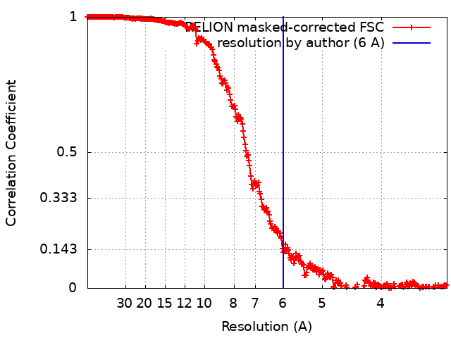
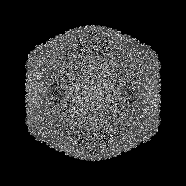
ジャーナル: Commun Biol / 年: 2022 タイトル: UVC inactivation of pathogenic samples suitable for cryo-EM analysis. 著者: Jamie S Depelteau / Ludovic Renault / Nynke Althof / C Keith Cassidy / Luiza M Mendonça / Grant J Jensen / Guenter P Resch / Ariane Briegel / 要旨: Cryo-electron microscopy has become an essential tool to understand structure and function of biological samples. Especially for pathogens, such as disease-causing bacteria and viruses, insights ...Cryo-electron microscopy has become an essential tool to understand structure and function of biological samples. Especially for pathogens, such as disease-causing bacteria and viruses, insights gained by cryo-EM can aid in developing cures. However, due to the biosafety restrictions of pathogens, samples are often treated by chemical fixation to render the pathogen inert, affecting the ultrastructure of the sample. Alternatively, researchers use in vitro or ex vivo models, which are non-pathogenic but lack the complexity of the pathogen of interest. Here we show that ultraviolet-C (UVC) radiation applied at cryogenic temperatures can be used to eliminate or dramatically reduce the infectivity of Vibrio cholerae and the bacterial virus, the ICP1 bacteriophage. We show no discernable structural impact of this treatment of either sample using two cryo-EM methods: cryo-electron tomography followed by sub-tomogram averaging, and single particle analysis (SPA). Additionally, we applied the UVC irradiation to the protein apoferritin (ApoF), which is a widely used test sample for high-resolution SPA studies. The UVC-treated ApoF sample resulted in a 2.1 Å structure indistinguishable from an untreated published map. This research demonstrates that UVC treatment is an effective and inexpensive addition to the cryo-EM sample preparation toolbox.
 ムービー
ムービー コントローラー


 データを開く
 基本情報
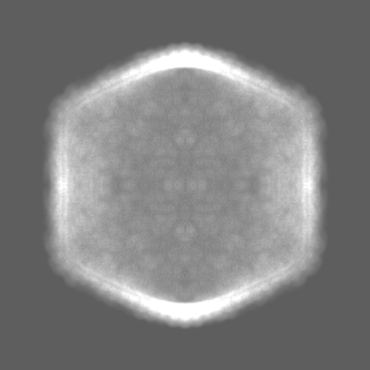
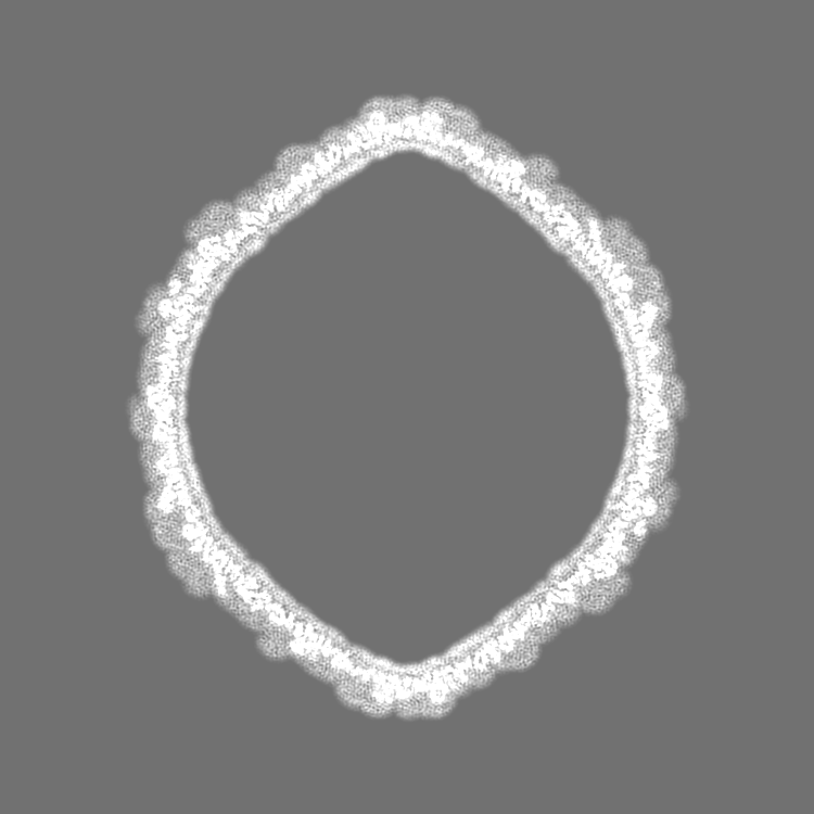
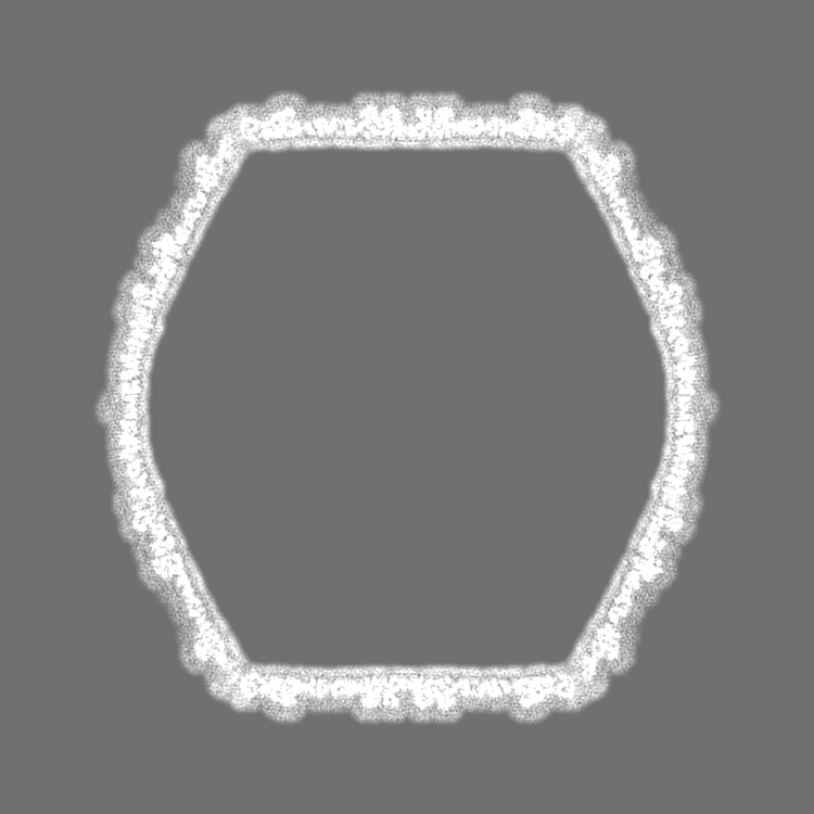
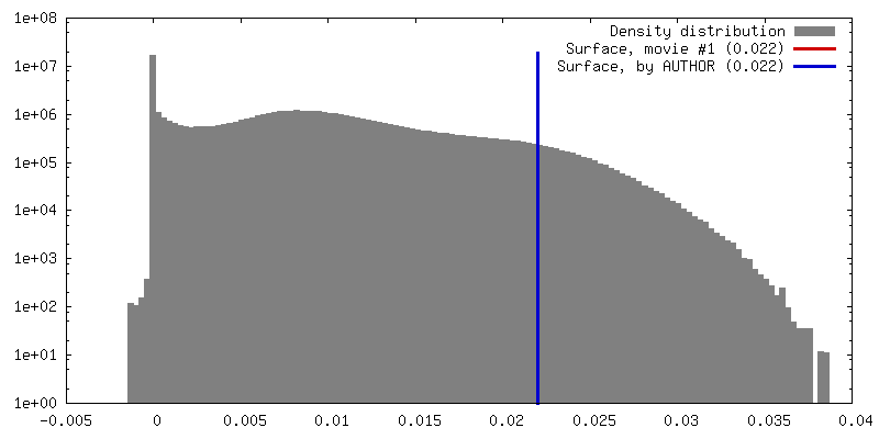
基本情報 マップデータ
 試料
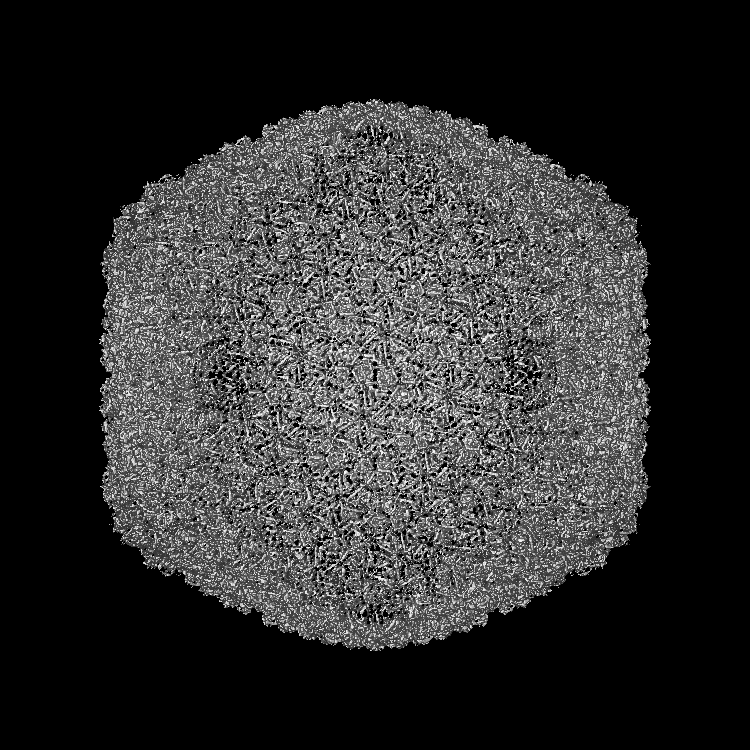
試料 Vibrio phage ICP1 (ファージ)
 データ登録者
 引用
引用


 構造の表示
構造の表示 ムービービューア
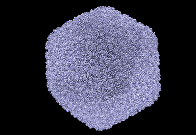
 ダウンロードとリンク
 emd_13403.png
 http://ftp.pdbj.org/pub/emdb/structures/EMD-13403

 Z (Sec.)
 Y (Row.)
 X (Col.)


 試料の構成要素
 Vibrio cholerae (コレラ菌) / 株: N16961
 解析
解析 電子顕微鏡法
 FIELD EMISSION GUN