 ムービー
ムービー コントローラー


+ データを開く

- 基本情報
基本情報
| 登録情報 |  | ||||||||||||
|---|---|---|---|---|---|---|---|---|---|---|---|---|---|
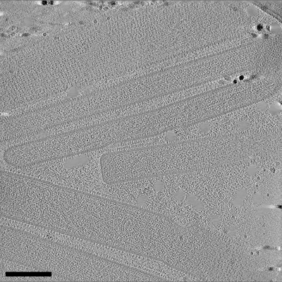
| タイトル | Cryo-electron tomogram of a pseudotype virus particle generated during coinfection of Influenza A virus and Respiratory Syncytial virus in lung cells. | ||||||||||||
 マップデータ マップデータ | Cryo-electron tomogram of a pseudotype virus particle generated during coinfection of Influenza A virus and Respiratory Syncytial virus in lung cells. | ||||||||||||
 試料 試料 | Pseudotype virus particle from RSV A2 and IAV PR8 != Respiratory syncytial virus A2 Pseudotype virus particle from RSV A2 and IAV PR8
| ||||||||||||
 キーワード キーワード | pseudotype virus particle / glycoprotein spikes / RNPs / VIRUS | ||||||||||||
| 生物種 |  Respiratory syncytial virus A2 (ウイルス) Respiratory syncytial virus A2 (ウイルス) | ||||||||||||
| 手法 | 電子線トモグラフィー法 / クライオ電子顕微鏡法 | ||||||||||||
 データ登録者 データ登録者 | Vijayakrishnan S / Murcia PR | ||||||||||||
| 資金援助 |  英国, 3件 英国, 3件
| ||||||||||||
 引用 引用 | ジャーナル: Nat Microbiol / 年: 2022 タイトル: Coinfection by influenza A virus and respiratory syncytial virus produces hybrid virus particles. 著者: Joanne Haney / Swetha Vijayakrishnan / James Streetley / Kieran Dee / Daniel Max Goldfarb / Mairi Clarke / Margaret Mullin / Stephen D Carter / David Bhella / Pablo R Murcia / 要旨: Interactions between respiratory viruses during infection affect transmission dynamics and clinical outcomes. To identify and characterize virus-virus interactions at the cellular level, we ...Interactions between respiratory viruses during infection affect transmission dynamics and clinical outcomes. To identify and characterize virus-virus interactions at the cellular level, we coinfected human lung cells with influenza A virus (IAV) and respiratory syncytial virus (RSV). Super-resolution microscopy, live-cell imaging, scanning electron microscopy and cryo-electron tomography revealed extracellular and membrane-associated filamentous structures consistent with hybrid viral particles (HVPs). We found that HVPs harbour surface glycoproteins and ribonucleoproteins of IAV and RSV. HVPs use the RSV fusion glycoprotein to evade anti-IAV neutralizing antibodies and infect and spread among cells lacking IAV receptors. Finally, we show that IAV and RSV coinfection in primary cells of the bronchial epithelium results in viral proteins from both viruses co-localizing at the apical cell surface. Our observations define a previously unknown interaction between respiratory viruses that might affect virus pathogenesis by expanding virus tropism and enabling immune evasion. | ||||||||||||
| 履歴 |
|
- 構造の表示
構造の表示
| 添付画像 |
|---|
- ダウンロードとリンク
-EMDBアーカイブ
| マップデータ | emd_13229.map.gz | 201 MB |  EMDBマップデータ形式 EMDBマップデータ形式 | |
|---|---|---|---|---|
| ヘッダ (付随情報) | emd-13229-v30.xmlemd-13229.xml | 11.2 KB 11.2 KB | 表示 表示 | EMDBヘッダ |
| 画像 |  emd_13229.png emd_13229.png | 244.7 KB | ||
| Filedesc metadata | emd-13229.cif.gz | 4.6 KB | ||
| アーカイブディレクトリ |  http://ftp.pdbj.org/pub/emdb/structures/EMD-13229ftp://ftp.pdbj.org/pub/emdb/structures/EMD-13229 http://ftp.pdbj.org/pub/emdb/structures/EMD-13229ftp://ftp.pdbj.org/pub/emdb/structures/EMD-13229 | HTTPS FTP |
-検証レポート
| 文書・要旨 | emd_13229_validation.pdf.gz | 449.6 KB | 表示 | EMDB検証レポート |
|---|---|---|---|---|
| 文書・詳細版 | emd_13229_full_validation.pdf.gz | 449.2 KB | 表示 | |
| XML形式データ | emd_13229_validation.xml.gz | 3.9 KB | 表示 | |
| CIF形式データ | emd_13229_validation.cif.gz | 4.5 KB | 表示 | |
| アーカイブディレクトリ | https://ftp.pdbj.org/pub/emdb/validation_reports/EMD-13229ftp://ftp.pdbj.org/pub/emdb/validation_reports/EMD-13229 | HTTPS FTP |
-関連構造データ

-リンク
| EMDBのページ | EMDB (EBI/PDBe) / EMDataResource |
|---|
-マップ
| ファイル | ダウンロード / ファイル: emd_13229.map.gz / 形式: CCP4 / 大きさ: 292 MB / タイプ: IMAGE STORED AS SIGNED INTEGER (2 BYTES) | ||||||||||||||||||||||||||||||||
|---|---|---|---|---|---|---|---|---|---|---|---|---|---|---|---|---|---|---|---|---|---|---|---|---|---|---|---|---|---|---|---|---|---|
| 注釈 | Cryo-electron tomogram of a pseudotype virus particle generated during coinfection of Influenza A virus and Respiratory Syncytial virus in lung cells. | ||||||||||||||||||||||||||||||||
| 投影像・断面図 | 画像のコントロール
画像は Spider により作成 これらの図は立方格子座標系で作成されたものです | ||||||||||||||||||||||||||||||||
| ボクセルのサイズ | X=Y=Z: 11.992 Å | ||||||||||||||||||||||||||||||||
| 密度 |
| ||||||||||||||||||||||||||||||||
| 対称性 | 空間群: 1 | ||||||||||||||||||||||||||||||||
| 詳細 | EMDB XML:
|
 Z (Sec.)
 Y (Row.)
 X (Col.)


-添付データ
- 試料の構成要素
-全体 : Pseudotype virus particle from RSV A2 and IAV PR8
| 全体 | 名称: Pseudotype virus particle from RSV A2 and IAV PR8 |
|---|---|
| 要素 |
|
-超分子 #1: Respiratory syncytial virus A2
| 超分子 | 名称: Respiratory syncytial virus A2 / タイプ: virus / ID: 1 / 親要素: 0 詳細: This sample was obtained by coinfection of A549 cells with both the PR8 strain of Influenza A virus and the A2 strain of Respiratory Syncytial Virus. NCBI-ID: 1972429 / 生物種: Respiratory syncytial virus A2 / Sci species strain: RSV A2 and IAV PR8 / ウイルスタイプ: VIRION / ウイルス・単離状態: STRAIN / ウイルス・エンベロープ: Yes / ウイルス・中空状態: No |
|---|---|
| 宿主 | 生物種:  Homo sapiens (ヒト) / 株: PR8 and A2 Homo sapiens (ヒト) / 株: PR8 and A2 |
-実験情報
-構造解析
| 手法 | クライオ電子顕微鏡法 |
|---|---|
 解析 解析 | 電子線トモグラフィー法 |
| 試料の集合状態 | cell |
-試料調製
| 緩衝液 | pH: 7.5 |
|---|---|
| グリッド | モデル: Quantifoil R2/2 / 材質: GOLD / メッシュ: 200 |
| 凍結 | 凍結剤: ETHANE / チャンバー内湿度: 95 % / チャンバー内温度: 277 K / 装置: FEI VITROBOT MARK IV 詳細: Blotted from backside for 7 seconds before plunging. |
| 詳細 | This sample was A549 lung fibroblast cells coinfected with the human PR8 strain of Influenza A virus (IAV) and the the A2 strain of Respiratory Syncytial virus (RSV) directly on EM grids prior to plunge freezing and imaging by Cryo-ET. |
| 切片作成 | その他: NO SECTIONING |
| 位置合わせマーカー | Manufacturer: British Biocell International / 直径: 20 nm |
- 電子顕微鏡法
| 顕微鏡 | JEOL CRYO ARM 300 |
|---|---|
| 特殊光学系 | エネルギーフィルター - 名称: In-column Omega Filter エネルギーフィルター - スリット幅: 30 eV |
| 撮影 | フィルム・検出器のモデル: DIRECT ELECTRON DE-64 (8k x 8k) 検出モード: COUNTING / 平均電子線量: 1.6 e/Å2 |
| 電子線 | 加速電圧: 300 kV / 電子線源:  FIELD EMISSION GUN FIELD EMISSION GUN |
| 電子光学系 | 照射モード: FLOOD BEAM / 撮影モード: BRIGHT FIELD / Cs: 2.7 mm / 最大 デフォーカス(公称値): 5.0 µm / 最小 デフォーカス(公称値): 4.0 µm / 倍率(公称値): 50000 |
| 試料ステージ | 試料ホルダーモデル: JEOL CRYOSPECPORTER / ホルダー冷却材: NITROGEN |
-画像解析
| 最終 再構成 | アルゴリズム: BACK PROJECTION / ソフトウェア - 名称: IMOD (ver. 4.11) ソフトウェア - 詳細: Tomograms reconstructed using Etomo module of IMOD and binned by 4 詳細: Raw images were aligned and dose corrected using alignframes module in IMOD to generate aligned tilt series. These were then used to reconstruct tomograms using weighted back projection via ...詳細: Raw images were aligned and dose corrected using alignframes module in IMOD to generate aligned tilt series. These were then used to reconstruct tomograms using weighted back projection via the stomp module in IMOD. CTF correction was carried out using the ctfplotter in IMOD prior to tomogram generation. Reconstructed tomograms were binned by 8 (bin8) to aid in visualisation and interpretation. 使用した粒子像数: 61 |
|---|
