 Movie
Movie Controller

[English] 日本語
 Yorodumi
- EMDB-13229: Cryo-electron tomogram of a pseudotype virus particle generated d... -
+ Open data

- Basic information
| Entry |  | ||||||||||||
|---|---|---|---|---|---|---|---|---|---|---|---|---|---|
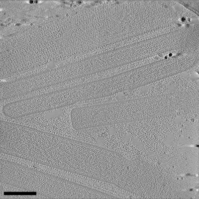
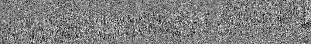
| Title | Cryo-electron tomogram of a pseudotype virus particle generated during coinfection of Influenza A virus and Respiratory Syncytial virus in lung cells. | ||||||||||||
 Map data Map data | Cryo-electron tomogram of a pseudotype virus particle generated during coinfection of Influenza A virus and Respiratory Syncytial virus in lung cells. | ||||||||||||
 Sample Sample | Pseudotype virus particle from RSV A2 and IAV PR8 != Respiratory syncytial virus A2 Pseudotype virus particle from RSV A2 and IAV PR8
| ||||||||||||
 Keywords Keywords | pseudotype virus particle / glycoprotein spikes / RNPs / VIRUS | ||||||||||||
| Biological species |  Respiratory syncytial virus A2 Respiratory syncytial virus A2 | ||||||||||||
| Method | electron tomography / cryo EM | ||||||||||||
 Authors Authors | Vijayakrishnan S / Murcia PR | ||||||||||||
| Funding support |  United Kingdom, 3 items United Kingdom, 3 items
| ||||||||||||
 Citation Citation | Journal: Nat Microbiol / Year: 2022 Title: Coinfection by influenza A virus and respiratory syncytial virus produces hybrid virus particles. Authors: Joanne Haney / Swetha Vijayakrishnan / James Streetley / Kieran Dee / Daniel Max Goldfarb / Mairi Clarke / Margaret Mullin / Stephen D Carter / David Bhella / Pablo R Murcia / Abstract: Interactions between respiratory viruses during infection affect transmission dynamics and clinical outcomes. To identify and characterize virus-virus interactions at the cellular level, we ...Interactions between respiratory viruses during infection affect transmission dynamics and clinical outcomes. To identify and characterize virus-virus interactions at the cellular level, we coinfected human lung cells with influenza A virus (IAV) and respiratory syncytial virus (RSV). Super-resolution microscopy, live-cell imaging, scanning electron microscopy and cryo-electron tomography revealed extracellular and membrane-associated filamentous structures consistent with hybrid viral particles (HVPs). We found that HVPs harbour surface glycoproteins and ribonucleoproteins of IAV and RSV. HVPs use the RSV fusion glycoprotein to evade anti-IAV neutralizing antibodies and infect and spread among cells lacking IAV receptors. Finally, we show that IAV and RSV coinfection in primary cells of the bronchial epithelium results in viral proteins from both viruses co-localizing at the apical cell surface. Our observations define a previously unknown interaction between respiratory viruses that might affect virus pathogenesis by expanding virus tropism and enabling immune evasion. | ||||||||||||
| History |
|
- Structure visualization
| Supplemental images |
|---|
- Downloads & links
-EMDB archive
| Map data | emd_13229.map.gz | 201 MB |  EMDB map data format EMDB map data format | |
|---|---|---|---|---|
| Header (meta data) | emd-13229-v30.xmlemd-13229.xml | 11.2 KB 11.2 KB | Display Display | EMDB header |
| Images |  emd_13229.png emd_13229.png | 244.7 KB | ||
| Filedesc metadata | emd-13229.cif.gz | 4.6 KB | ||
| Archive directory |  http://ftp.pdbj.org/pub/emdb/structures/EMD-13229ftp://ftp.pdbj.org/pub/emdb/structures/EMD-13229 http://ftp.pdbj.org/pub/emdb/structures/EMD-13229ftp://ftp.pdbj.org/pub/emdb/structures/EMD-13229 | HTTPS FTP |
-Related structure data

-Links
| EMDB pages | EMDB (EBI/PDBe) / EMDataResource |
|---|
-Map
| File | Download / File: emd_13229.map.gz / Format: CCP4 / Size: 292 MB / Type: IMAGE STORED AS SIGNED INTEGER (2 BYTES) | ||||||||||||||||||||||||||||||||
|---|---|---|---|---|---|---|---|---|---|---|---|---|---|---|---|---|---|---|---|---|---|---|---|---|---|---|---|---|---|---|---|---|---|
| Annotation | Cryo-electron tomogram of a pseudotype virus particle generated during coinfection of Influenza A virus and Respiratory Syncytial virus in lung cells. | ||||||||||||||||||||||||||||||||
| Projections & slices | Image control
Images are generated by Spider. generated in cubic-lattice coordinate | ||||||||||||||||||||||||||||||||
| Voxel size | X=Y=Z: 11.992 Å | ||||||||||||||||||||||||||||||||
| Density |
| ||||||||||||||||||||||||||||||||
| Symmetry | Space group: 1 | ||||||||||||||||||||||||||||||||
| Details | EMDB XML:
|
 Z (Sec.)
 Y (Row.)
 X (Col.)


-Supplemental data
- Sample components
-Entire : Pseudotype virus particle from RSV A2 and IAV PR8
| Entire | Name: Pseudotype virus particle from RSV A2 and IAV PR8 |
|---|---|
| Components |
|
-Supramolecule #1: Respiratory syncytial virus A2
| Supramolecule | Name: Respiratory syncytial virus A2 / type: virus / ID: 1 / Parent: 0 Details: This sample was obtained by coinfection of A549 cells with both the PR8 strain of Influenza A virus and the A2 strain of Respiratory Syncytial Virus. NCBI-ID: 1972429 / Sci species name: Respiratory syncytial virus A2 / Sci species strain: RSV A2 and IAV PR8 / Virus type: VIRION / Virus isolate: STRAIN / Virus enveloped: Yes / Virus empty: No |
|---|---|
| Host (natural) | Organism:  Homo sapiens (human) / Strain: PR8 and A2 Homo sapiens (human) / Strain: PR8 and A2 |
-Experimental details
-Structure determination
| Method | cryo EM |
|---|---|
 Processing Processing | electron tomography |
| Aggregation state | cell |
-Sample preparation
| Buffer | pH: 7.5 |
|---|---|
| Grid | Model: Quantifoil R2/2 / Material: GOLD / Mesh: 200 |
| Vitrification | Cryogen name: ETHANE / Chamber humidity: 95 % / Chamber temperature: 277 K / Instrument: FEI VITROBOT MARK IV Details: Blotted from backside for 7 seconds before plunging. |
| Details | This sample was A549 lung fibroblast cells coinfected with the human PR8 strain of Influenza A virus (IAV) and the the A2 strain of Respiratory Syncytial virus (RSV) directly on EM grids prior to plunge freezing and imaging by Cryo-ET. |
| Sectioning | Other: NO SECTIONING |
| Fiducial marker | Manufacturer: British Biocell International / Diameter: 20 nm |
- Electron microscopy
| Microscope | JEOL CRYO ARM 300 |
|---|---|
| Specialist optics | Energy filter - Name: In-column Omega Filter / Energy filter - Slit width: 30 eV |
| Image recording | Film or detector model: DIRECT ELECTRON DE-64 (8k x 8k) / Detector mode: COUNTING / Average electron dose: 1.6 e/Å2 |
| Electron beam | Acceleration voltage: 300 kV / Electron source:  FIELD EMISSION GUN FIELD EMISSION GUN |
| Electron optics | Illumination mode: FLOOD BEAM / Imaging mode: BRIGHT FIELD / Cs: 2.7 mm / Nominal defocus max: 5.0 µm / Nominal defocus min: 4.0 µm / Nominal magnification: 50000 |
| Sample stage | Specimen holder model: JEOL CRYOSPECPORTER / Cooling holder cryogen: NITROGEN |
-Image processing
| Final reconstruction | Algorithm: BACK PROJECTION / Software - Name: IMOD (ver. 4.11) Software - details: Tomograms reconstructed using Etomo module of IMOD and binned by 4 Details: Raw images were aligned and dose corrected using alignframes module in IMOD to generate aligned tilt series. These were then used to reconstruct tomograms using weighted back projection via ...Details: Raw images were aligned and dose corrected using alignframes module in IMOD to generate aligned tilt series. These were then used to reconstruct tomograms using weighted back projection via the stomp module in IMOD. CTF correction was carried out using the ctfplotter in IMOD prior to tomogram generation. Reconstructed tomograms were binned by 8 (bin8) to aid in visualisation and interpretation. Number images used: 61 |
|---|
