 ムービー
ムービー コントローラー


+ データを開く

- 基本情報
基本情報
| 登録情報 | データベース: EMDB / ID: EMD-1320 | |||||||||
|---|---|---|---|---|---|---|---|---|---|---|
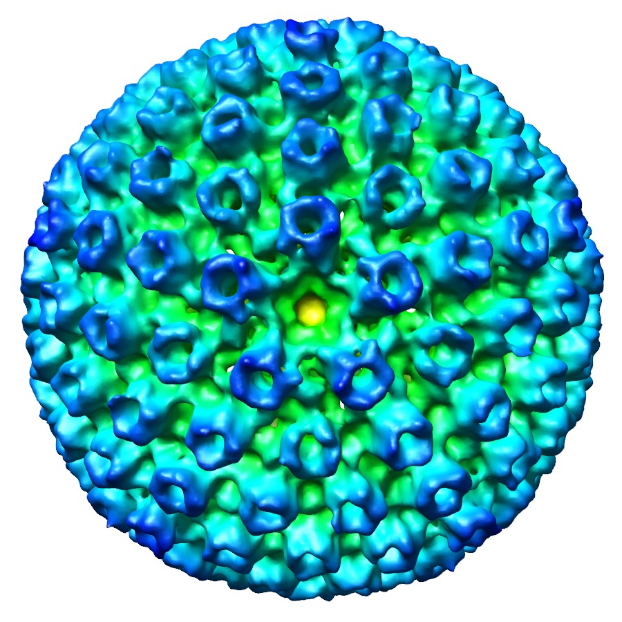
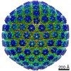
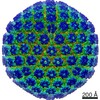
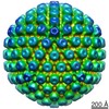
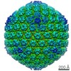
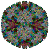
| タイトル | Direct visualization of the putative portal in the Kaposi's sarcoma-associated herpesvirus capsid by cryoelectron tomography. | |||||||||
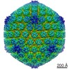
 マップデータ マップデータ | This is a MRC density map averaged from KSHV A-capsid tomograms. 5-fold symmetry is applied along the unique vertex. | |||||||||
 試料 試料 |
| |||||||||
| 生物種 |   Human herpesvirus 8 (ヘルペスウイルス) Human herpesvirus 8 (ヘルペスウイルス) | |||||||||
| 手法 | サブトモグラム平均法 / クライオ電子顕微鏡法 / 解像度: 40.0 Å | |||||||||
 データ登録者 データ登録者 | Deng B / O'Connor CM / Kedes DH / Zhou ZH | |||||||||
 引用 引用 | ジャーナル: J Virol / 年: 2007 タイトル: Direct visualization of the putative portal in the Kaposi's sarcoma-associated herpesvirus capsid by cryoelectron tomography. 著者: Binbin Deng / Christine M O'Connor / Dean H Kedes / Z Hong Zhou /  要旨: Genetic and biochemical studies have suggested the existence of a bacteriophage-like, DNA-packaging/ejecting portal complex in herpesviruses capsids, but its arrangement remained unknown. Here, we ...Genetic and biochemical studies have suggested the existence of a bacteriophage-like, DNA-packaging/ejecting portal complex in herpesviruses capsids, but its arrangement remained unknown. Here, we report the first visualization of a unique vertex in the Kaposi's sarcoma-associated herpesvirus (KSHV) capsid by cryoelectron tomography, thus providing direct structural evidence for the existence of a portal complex in a gammaherpesvirus. This putative KSHV portal is an internally localized, umbilicated structure and lacks all of the external machineries characteristic of portals in DNA bacteriophages. | |||||||||
| 履歴 |
|
- 構造の表示
構造の表示
| ムービー |
 ムービービューア ムービービューア |
|---|---|
| 構造ビューア | EMマップ: SurfViewMolmilJmol/JSmol |
| 添付画像 |


- ダウンロードとリンク
-EMDBアーカイブ
| マップデータ | emd_1320.map.gz | 16.7 MB | EMDBマップデータ形式 | |
|---|---|---|---|---|
| ヘッダ (付随情報) | emd-1320-v30.xmlemd-1320.xml | 8.9 KB 8.9 KB | 表示 表示 | EMDBヘッダ |
| 画像 | 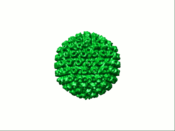 1320.gifemd_1320.tifemd_1320_1.tifemd_1320_2.tifemd_1320_3.tif 1320.gifemd_1320.tifemd_1320_1.tifemd_1320_2.tifemd_1320_3.tif | 46.1 KB 2.3 MB 2.3 MB 2.3 MB 2.3 MB | ||
| アーカイブディレクトリ |  http://ftp.pdbj.org/pub/emdb/structures/EMD-1320ftp://ftp.pdbj.org/pub/emdb/structures/EMD-1320 http://ftp.pdbj.org/pub/emdb/structures/EMD-1320ftp://ftp.pdbj.org/pub/emdb/structures/EMD-1320 | HTTPS FTP |
-関連構造データ

-リンク
| EMDBのページ | EMDB (EBI/PDBe) / EMDataResource |
|---|
-マップ
| ファイル | ダウンロード / ファイル: emd_1320.map.gz / 形式: CCP4 / 大きさ: 17.7 MB / タイプ: IMAGE STORED AS FLOATING POINT NUMBER (4 BYTES) | ||||||||||||||||||||||||||||||||||||||||||||||||||||||||||||||||||||
|---|---|---|---|---|---|---|---|---|---|---|---|---|---|---|---|---|---|---|---|---|---|---|---|---|---|---|---|---|---|---|---|---|---|---|---|---|---|---|---|---|---|---|---|---|---|---|---|---|---|---|---|---|---|---|---|---|---|---|---|---|---|---|---|---|---|---|---|---|---|
| 注釈 | This is a MRC density map averaged from KSHV A-capsid tomograms. 5-fold symmetry is applied along the unique vertex. | ||||||||||||||||||||||||||||||||||||||||||||||||||||||||||||||||||||
| 投影像・断面図 | 画像のコントロール
画像は Spider により作成 | ||||||||||||||||||||||||||||||||||||||||||||||||||||||||||||||||||||
| ボクセルのサイズ | X=Y=Z: 7.858 Å | ||||||||||||||||||||||||||||||||||||||||||||||||||||||||||||||||||||
| 密度 |
| ||||||||||||||||||||||||||||||||||||||||||||||||||||||||||||||||||||
| 対称性 | 空間群: 1 | ||||||||||||||||||||||||||||||||||||||||||||||||||||||||||||||||||||
| 詳細 | EMDB XML:
CCP4マップ ヘッダ情報:
| ||||||||||||||||||||||||||||||||||||||||||||||||||||||||||||||||||||
 Z (Sec.)
 Y (Row.)
 X (Col.)


-添付データ
- 試料の構成要素
-全体 : KSHV
| 全体 | 名称: KSHV |
|---|---|
| 要素 |
|
-超分子 #1000: KSHV
| 超分子 | 名称: KSHV / タイプ: sample / ID: 1000 / 集合状態: 1 / Number unique components: 1 |
|---|
-超分子 #1: Human herpesvirus 8
| 超分子 | 名称: Human herpesvirus 8 / タイプ: virus / ID: 1 / Name.synonym: Kaposis sarcoma-associated herpesvirus / NCBI-ID: 37296 / 生物種: Human herpesvirus 8 / データベース: NCBI / ウイルスタイプ: VIRION / ウイルス・単離状態: STRAIN / ウイルス・エンベロープ: No / ウイルス・中空状態: Yes / Syn species name: Kaposis sarcoma-associated herpesvirus |
|---|---|
| 宿主 | 生物種:  Homo sapiens (ヒト) / 別称: VERTEBRATES Homo sapiens (ヒト) / 別称: VERTEBRATES |
| ウイルス殻 | Shell ID: 1 / 直径: 1250 Å / T番号(三角分割数): 16 |
-実験情報
-構造解析
| 手法 | クライオ電子顕微鏡法 |
|---|---|
 解析 解析 | サブトモグラム平均法 |
| 試料の集合状態 | particle |
-試料調製
| 緩衝液 | pH: 7.4 / 詳細: PBS |
|---|---|
| グリッド | 詳細: holey carbon 300 mesh copper grid |
| 凍結 | 凍結剤: ETHANE / 装置: HOMEMADE PLUNGER / 詳細: Vitrification instrument: plunger / 手法: Blot for 1 seconds before plunging |
- 電子顕微鏡法
| 顕微鏡 | FEI TECNAI F30 |
|---|---|
| 温度 | 最低: 90 K / 最高: 90 K / 平均: 90 K |
| 詳細 | Tilted from -70 to +70 with 2 degree interval. |
| 撮影 | カテゴリ: CCD / フィルム・検出器のモデル: GENERIC CCD / 平均電子線量: 1 e/Å2 |
| 電子線 | 加速電圧: 300 kV / 電子線源:  FIELD EMISSION GUN FIELD EMISSION GUN |
| 電子光学系 | 倍率(補正後): 38200 / 照射モード: FLOOD BEAM / 撮影モード: BRIGHT FIELD / Cs: 2 mm / 最大 デフォーカス(公称値): 8.0 µm / 最小 デフォーカス(公称値): 6.0 µm / 倍率(公称値): 23000 |
| 試料ステージ | 試料ホルダー: Side entry liquid nitrogen-cooled cryo specimen holder 試料ホルダーモデル: GATAN LIQUID NITROGEN / Tilt series - Axis1 - Min angle: 70 ° / Tilt series - Axis1 - Max angle: 70 ° |
| 実験機器 |  モデル: Tecnai F30 / 画像提供: FEI Company |
-画像解析
| 最終 再構成 | アルゴリズム: OTHER / 解像度のタイプ: BY AUTHOR / 解像度: 40.0 Å / 解像度の算出法: OTHER / ソフトウェア - 名称: PROJALIGN |
|---|
