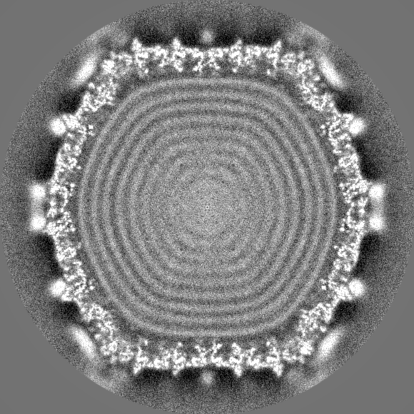
Journal: Nature / Year: 2004 Title: Membrane structure and interactions with protein and DNA in bacteriophage PRD1. Authors: Joseph J B Cockburn / Nicola G A Abrescia / Jonathan M Grimes / Geoffrey C Sutton / Jonathan M Diprose / James M Benevides / George J Thomas / Jaana K H Bamford / Dennis H Bamford / David I Stuart / Abstract: Membranes are essential for selectively controlling the passage of molecules in and out of cells and mediating the response of cells to their environment. Biological membranes and their associated ...Membranes are essential for selectively controlling the passage of molecules in and out of cells and mediating the response of cells to their environment. Biological membranes and their associated proteins present considerable difficulties for structural analysis. Although enveloped viruses have been imaged at about 9 A resolution by cryo-electron microscopy and image reconstruction, no detailed crystallographic structure of a membrane system has been described. The structure of the bacteriophage PRD1 particle, determined by X-ray crystallography at about 4 A resolution, allows the first detailed analysis of a membrane-containing virus. The architecture of the viral capsid and its implications for virus assembly are presented in the accompanying paper. Here we show that the electron density also reveals the icosahedral lipid bilayer, beneath the protein capsid, enveloping the viral DNA. The viral membrane contains about 26,000 lipid molecules asymmetrically distributed between the membrane leaflets. The inner leaflet is composed predominantly of zwitterionic phosphatidylethanolamine molecules, facilitating a very close interaction with the viral DNA, which we estimate to be packaged to a pressure of about 45 atm, factors that are likely to be important during membrane-mediated DNA translocation into the host cell. In contrast, the outer leaflet is enriched in phosphatidylglycerol and cardiolipin, which show a marked lateral segregation within the icosahedral asymmetric unit. In addition, the lipid headgroups show a surprising degree of order.
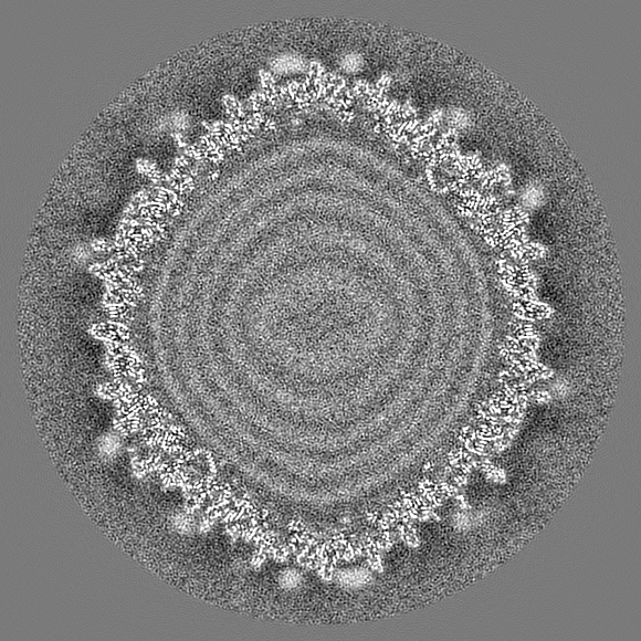
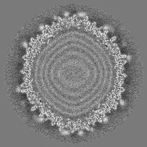
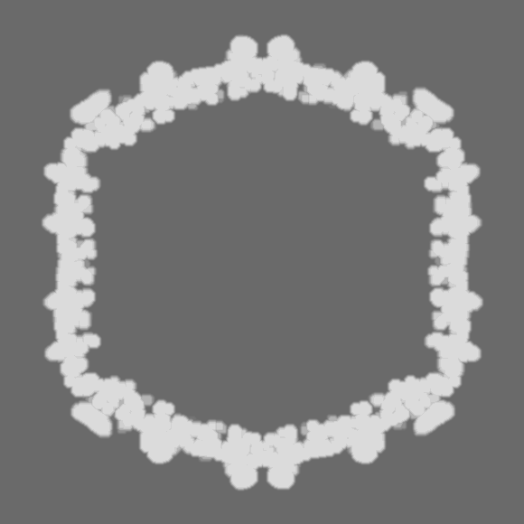
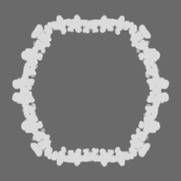
the virus sample was mixed with CPZ and different concetration on the grid were explored
-
Electron microscopy
Microscope
FEI TITAN KRIOS
Image recording
Film or detector model: FEI FALCON III (4k x 4k) / Detector mode: INTEGRATING / Number grids imaged: 2 / Number real images: 3218 / Average electron dose: 36.0 e/Å2
Electron beam
Acceleration voltage: 300 kV / Electron source: FIELD EMISSION GUN
Electron optics
C2 aperture diameter: 50.0 µm / Illumination mode: OTHER / Imaging mode: BRIGHT FIELD / Cs: 2.7 mm
In the structure databanks used in Yorodumi, some data are registered as the other names, "COVID-19 virus" and "2019-nCoV". Here are the details of the virus and the list of structure data.
Jan 31, 2019. EMDB accession codes are about to change! (news from PDBe EMDB page)
EMDB accession codes are about to change! (news from PDBe EMDB page)
The allocation of 4 digits for EMDB accession codes will soon come to an end. Whilst these codes will remain in use, new EMDB accession codes will include an additional digit and will expand incrementally as the available range of codes is exhausted. The current 4-digit format prefixed with “EMD-” (i.e. EMD-XXXX) will advance to a 5-digit format (i.e. EMD-XXXXX), and so on. It is currently estimated that the 4-digit codes will be depleted around Spring 2019, at which point the 5-digit format will come into force.
The EM Navigator/Yorodumi systems omit the EMD- prefix.
Related info.:Q: What is EMD? / ID/Accession-code notation in Yorodumi/EM Navigator
Yorodumi is a browser for structure data from EMDB, PDB, SASBDB, etc.
This page is also the successor to EM Navigator detail page, and also detail information page/front-end page for Omokage search.
The word "yorodu" (or yorozu) is an old Japanese word meaning "ten thousand". "mi" (miru) is to see.
Related info.:EMDB / PDB / SASBDB / Comparison of 3 databanks / Yorodumi Search / Aug 31, 2016. New EM Navigator & Yorodumi / Yorodumi Papers / Jmol/JSmol / Function and homology information / Changes in new EM Navigator and Yorodumi
 Movie
Movie Controller


 Open data
 Basic information
 Map data
 Sample
 Enterobacteria phage PRD1 (virus)
 Authors
 Spain,
  United Kingdom, 5 items
  Citation
 Structure visualization
 Downloads & links
 EMDB map data format
 emd_13109.png
 http://ftp.pdbj.org/pub/emdb/structures/EMD-13109
 Z (Sec.)
 Y (Row.)
 X (Col.)


 Sample components
 Salmonella enterica (bacteria)
 Processing
 Electron microscopy
 FIELD EMISSION GUN