ムービー
ムービー コントローラー


+ データを開く

- 基本情報
基本情報
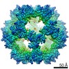
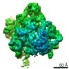
| 登録情報 | データベース: EMDB / ID: EMD-13093 | |||||||||
|---|---|---|---|---|---|---|---|---|---|---|
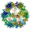
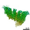
| タイトル | Immature 60S Ribosomal Subunit from C. thermophilum | |||||||||
 マップデータ マップデータ | Immature 60S Ribosomal Subunit from C.thermophilum, Main Map | |||||||||
 試料 試料 |
| |||||||||
| 生物種 |  Chaetomium thermophilum var. thermophilum DSM 1495 (菌類) Chaetomium thermophilum var. thermophilum DSM 1495 (菌類) | |||||||||
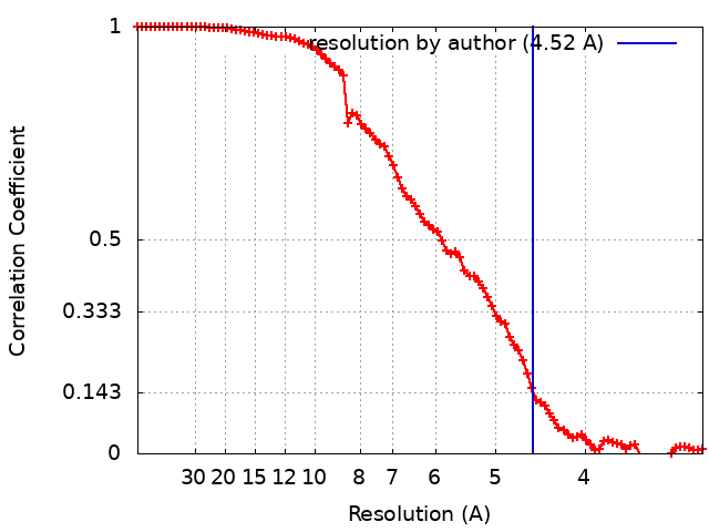
| 手法 | 単粒子再構成法 / クライオ電子顕微鏡法 / 解像度: 4.52 Å | |||||||||
 データ登録者 データ登録者 | Skalidis I / Kastritis PL | |||||||||
 引用 引用 | ジャーナル: Structure / 年: 2022 タイトル: Cryo-EM and artificial intelligence visualize endogenous protein community members. 著者: Ioannis Skalidis / Fotis L Kyrilis / Christian Tüting / Farzad Hamdi / Grzegorz Chojnowski / Panagiotis L Kastritis /  要旨: Cellular function is underlined by megadalton assemblies organizing in proximity, forming communities. Metabolons are protein communities involving metabolic pathways such as protein, fatty acid, and ...Cellular function is underlined by megadalton assemblies organizing in proximity, forming communities. Metabolons are protein communities involving metabolic pathways such as protein, fatty acid, and thioesters of coenzyme-A synthesis. Metabolons are highly heterogeneous due to their function, making their analysis particularly challenging. Here, we simultaneously characterize metabolon-embedded architectures of a 60S pre-ribosome, fatty acid synthase, and pyruvate/oxoglutarate dehydrogenase complex E2 cores de novo. Cryo-electron microscopy (cryo-EM) 3D reconstructions are resolved at 3.84-4.52 Å resolution by collecting <3,000 micrographs of a single cellular fraction. After combining cryo-EM with artificial intelligence-based atomic modeling and de novo sequence identification methods, at this resolution range, polypeptide hydrogen bonding patterns are discernible. Residing molecular components resemble their purified counterparts from other eukaryotes but also exhibit substantial conformational variation with potential functional implications. Our results propose an integrated tool, boosted by machine learning, that opens doors for structural systems biology spearheaded by cryo-EM characterization of native cell extracts. | |||||||||
| 履歴 |
|
- 構造の表示
構造の表示
| ムービー |
 ムービービューア ムービービューア |
|---|---|
| 構造ビューア | EMマップ: SurfViewMolmilJmol/JSmol |
| 添付画像 |

- ダウンロードとリンク
-EMDBアーカイブ
| マップデータ | emd_13093.map.gz | 59.6 MB | EMDBマップデータ形式 | |
|---|---|---|---|---|
| ヘッダ (付随情報) | emd-13093-v30.xmlemd-13093.xml | 31.4 KB 31.4 KB | 表示 表示 | EMDBヘッダ |
| FSC (解像度算出) | emd_13093_fsc.xml | 8.9 KB | 表示 | FSCデータファイル |
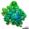
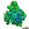
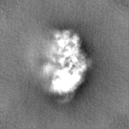
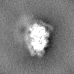
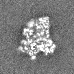
| 画像 | 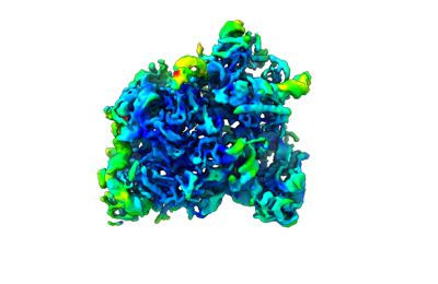 emd_13093.png emd_13093.png | 61.1 KB | ||
| その他 | emd_13093_additional_1.map.gzemd_13093_half_map_1.map.gzemd_13093_half_map_2.map.gz | 117.2 MB 59.5 MB 59.5 MB | ||
| アーカイブディレクトリ |  http://ftp.pdbj.org/pub/emdb/structures/EMD-13093ftp://ftp.pdbj.org/pub/emdb/structures/EMD-13093 http://ftp.pdbj.org/pub/emdb/structures/EMD-13093ftp://ftp.pdbj.org/pub/emdb/structures/EMD-13093 | HTTPS FTP |
-検証レポート
| 文書・要旨 | emd_13093_validation.pdf.gz | 529.7 KB | 表示 | EMDB検証レポート |
|---|---|---|---|---|
| 文書・詳細版 | emd_13093_full_validation.pdf.gz | 529.3 KB | 表示 | |
| XML形式データ | emd_13093_validation.xml.gz | 15.9 KB | 表示 | |
| CIF形式データ | emd_13093_validation.cif.gz | 20.5 KB | 表示 | |
| アーカイブディレクトリ | https://ftp.pdbj.org/pub/emdb/validation_reports/EMD-13093ftp://ftp.pdbj.org/pub/emdb/validation_reports/EMD-13093 | HTTPS FTP |
-関連構造データ
| 関連構造データ |  7q5qC 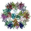 7q5rC  7q5sC C: 同じ文献を引用 ( |
|---|---|
| 類似構造データ | |
| 電子顕微鏡画像生データ | EMPIAR-10892 (タイトル: Cryo-EM SPA dataset of Megadalton-range protein communities from a Chaetomium thermophilum native cell extract Data size: 1.1 TB Data #1: Unaligned fractions saved by Falcon 3 EC camera [micrographs - multiframe]) |


-リンク
| EMDBのページ | EMDB (EBI/PDBe) / EMDataResource |
|---|---|
| 「今月の分子」の関連する項目 |

-マップ
| ファイル | ダウンロード / ファイル: emd_13093.map.gz / 形式: CCP4 / 大きさ: 64 MB / タイプ: IMAGE STORED AS FLOATING POINT NUMBER (4 BYTES) | ||||||||||||||||||||||||||||||||||||||||||||||||||||||||||||
|---|---|---|---|---|---|---|---|---|---|---|---|---|---|---|---|---|---|---|---|---|---|---|---|---|---|---|---|---|---|---|---|---|---|---|---|---|---|---|---|---|---|---|---|---|---|---|---|---|---|---|---|---|---|---|---|---|---|---|---|---|---|
| 注釈 | Immature 60S Ribosomal Subunit from C.thermophilum, Main Map | ||||||||||||||||||||||||||||||||||||||||||||||||||||||||||||
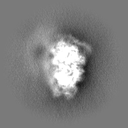
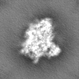
| 投影像・断面図 | 画像のコントロール
画像は Spider により作成 | ||||||||||||||||||||||||||||||||||||||||||||||||||||||||||||
| ボクセルのサイズ | X=Y=Z: 1.5678 Å | ||||||||||||||||||||||||||||||||||||||||||||||||||||||||||||
| 密度 |
| ||||||||||||||||||||||||||||||||||||||||||||||||||||||||||||
| 対称性 | 空間群: 1 | ||||||||||||||||||||||||||||||||||||||||||||||||||||||||||||
| 詳細 | EMDB XML:
CCP4マップ ヘッダ情報:
| ||||||||||||||||||||||||||||||||||||||||||||||||||||||||||||
 Z (Sec.)
 Y (Row.)
 X (Col.)


-添付データ
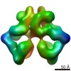
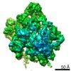
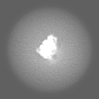
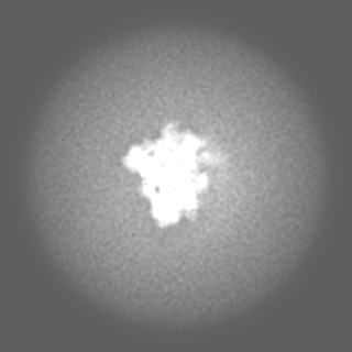
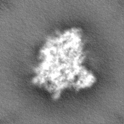
-追加マップ: Signature 4 Map
| ファイル | emd_13093_additional_1.map | ||||||||||||
|---|---|---|---|---|---|---|---|---|---|---|---|---|---|
| 注釈 | Signature 4 Map | ||||||||||||
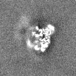
| 投影像・断面図 |
| ||||||||||||
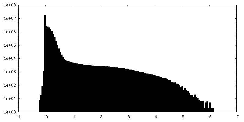
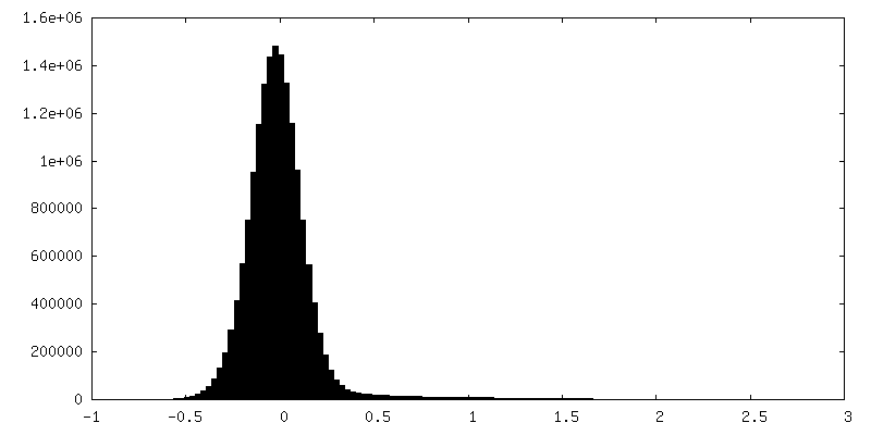
| 密度ヒストグラム |
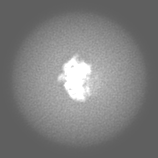
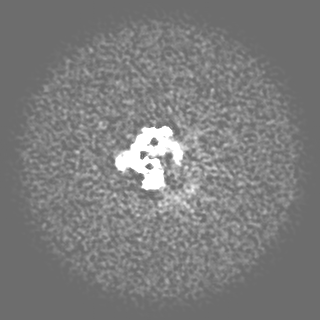


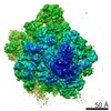
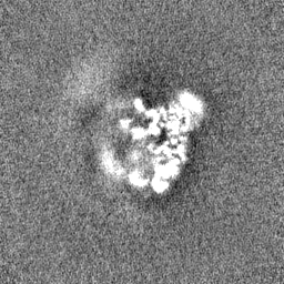
-ハーフマップ: Immature 60S Ribosomal Subunit from C.thermophilum, Half-Map A
| ファイル | emd_13093_half_map_1.map | ||||||||||||
|---|---|---|---|---|---|---|---|---|---|---|---|---|---|
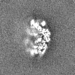
| 注釈 | Immature 60S Ribosomal Subunit from C.thermophilum, Half-Map A | ||||||||||||
| 投影像・断面図 |
| ||||||||||||
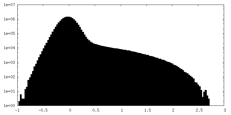
| 密度ヒストグラム |


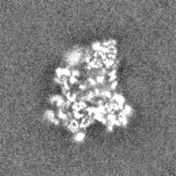


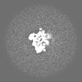
-ハーフマップ: Immature 60S Ribosomal Subunit from C.thermophilum, Half-Map B
| ファイル | emd_13093_half_map_2.map | ||||||||||||
|---|---|---|---|---|---|---|---|---|---|---|---|---|---|
| 注釈 | Immature 60S Ribosomal Subunit from C.thermophilum, Half-Map B | ||||||||||||
| 投影像・断面図 |
| ||||||||||||
| 密度ヒストグラム |

- 試料の構成要素
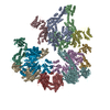
+全体 : Native 60-mer core of Pyruvate Dehydrogenase Complex
+超分子 #1: Native 60-mer core of Pyruvate Dehydrogenase Complex
+分子 #1: uL13
+分子 #2: eL21
+分子 #3: eL13
+分子 #4: eL15
+分子 #5: eL18
+分子 #6: uL15
+分子 #7: eL36
+分子 #8: eL32
+分子 #9: eL20
+分子 #10: uL22
+分子 #11: eL8
+分子 #12: uL29
+分子 #13: uL6
+分子 #14: eL6
+分子 #15: uL5
+分子 #16: uL18
+分子 #17: eL14
+分子 #18: uL30
+分子 #19: eL33
+分子 #20: uL24
+分子 #21: uL4
-実験情報
-構造解析
| 手法 | クライオ電子顕微鏡法 |
|---|---|
 解析 解析 | 単粒子再構成法 |
| 試料の集合状態 | particle |
-試料調製
| 濃度 | 0.3 mg/mL |
|---|---|
| 緩衝液 | pH: 7.4 / 構成要素 - 濃度: 200.0 mM / 構成要素 - 式: NH4CH2COOH / 構成要素 - 名称: Ammonium acetate |
| 凍結 | 凍結剤: ETHANE / チャンバー内湿度: 95 % / チャンバー内温度: 277 K / 装置: FEI VITROBOT MARK IV 詳細: For plunging, blot force 0 and blotting time of 4 sec were applied.. |
- 電子顕微鏡法
| 顕微鏡 | TFS GLACIOS |
|---|---|
| 温度 | 最低: 77.15 K / 最高: 103.15 K |
| アライメント法 | Coma free - Residual tilt: 14.7 mrad |
| 撮影 | フィルム・検出器のモデル: FEI FALCON III (4k x 4k) 検出モード: INTEGRATING / 実像数: 2808 / 平均電子線量: 30.0 e/Å2 |
| 電子線 | 加速電圧: 200 kV / 電子線源:  FIELD EMISSION GUN FIELD EMISSION GUN |
| 電子光学系 | C2レンズ絞り径: 100.0 µm / 照射モード: OTHER / 撮影モード: BRIGHT FIELD / Cs: 2.7 mm / 最大 デフォーカス(公称値): 2.0 µm / 最小 デフォーカス(公称値): 0.8 µm / 倍率(公称値): 92000 |
| 試料ステージ | 試料ホルダーモデル: OTHER / ホルダー冷却材: NITROGEN |