[English] 日本語
 Yorodumi
- EMDB-1296: An expanded conformation of single-ring GroEL-GroES complex encap... -
+ Open data
- Basic information
| Entry | Database: EMDB / ID: EMD-1296 | |||||||||
|---|---|---|---|---|---|---|---|---|---|---|
| Title | An expanded conformation of single-ring GroEL-GroES complex encapsulates an 86 kDa substrate. | |||||||||
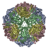
 Map data Map data | This is the surface representation of the electron density map at ~20-Angstrom resolution for the empty expanded conformation of SR398-AlphaBeta-GroES-Mg-ATP complex | |||||||||
 Sample Sample |
| |||||||||
| Biological species |  | |||||||||
| Method | single particle reconstruction / cryo EM / Resolution: 20.0 Å | |||||||||
 Authors Authors | Chen D-H / Song J-L / Chuang DT / Chiu W / Ludtke SJ | |||||||||
 Citation Citation |  Journal: Structure / Year: 2006 Journal: Structure / Year: 2006Title: An expanded conformation of single-ring GroEL-GroES complex encapsulates an 86 kDa substrate. Authors: Dong-Hua Chen / Jiu-Li Song / David T Chuang / Wah Chiu / Steven J Ludtke /  Abstract: Electron cryomicroscopy reveals an unprecedented conformation of the single-ring mutant of GroEL (SR398) bound to GroES in the presence of Mg-ATP. This conformation exhibits a considerable expansion ...Electron cryomicroscopy reveals an unprecedented conformation of the single-ring mutant of GroEL (SR398) bound to GroES in the presence of Mg-ATP. This conformation exhibits a considerable expansion of the folding cavity, with approximately 80% more volume than the X-ray structure of the equivalent cis cavity in the GroEL-GroES-(ADP)(7) complex. This expanded conformation can encapsulate an 86 kDa heterodimeric (alphabeta) assembly intermediate of mitochondrial branched-chain alpha-ketoacid dehydrogenase, the largest substrate ever observed to be cis encapsulated. The SR398-GroES-Mg-ATP complex is found to exist as a mixture of standard and expanded conformations, regardless of the absence or presence of the substrate. However, the presence of even a small substrate causes a pronounced bias toward the expanded conformation. Encapsulation of the large assembly intermediate is supported by a series of electron cryomicroscopy studies as well as the protection of both alpha and beta subunits of the substrate from tryptic digestion. | |||||||||
| History |
|
- Structure visualization
| Movie |
 Movie viewer Movie viewer |
|---|---|
| Structure viewer | EM map:  SurfView SurfView Molmil Molmil Jmol/JSmol Jmol/JSmol |
| Supplemental images |
- Downloads & links
-EMDB archive
| Map data |  emd_1296.map.gz emd_1296.map.gz | 4.2 MB |  EMDB map data format EMDB map data format | |
|---|---|---|---|---|
| Header (meta data) |  emd-1296-v30.xml emd-1296-v30.xml emd-1296.xml emd-1296.xml | 12 KB 12 KB | Display Display |  EMDB header EMDB header |
| Images |  1296.gif 1296.gif | 37.5 KB | ||
| Archive directory |  http://ftp.pdbj.org/pub/emdb/structures/EMD-1296 http://ftp.pdbj.org/pub/emdb/structures/EMD-1296 ftp://ftp.pdbj.org/pub/emdb/structures/EMD-1296 ftp://ftp.pdbj.org/pub/emdb/structures/EMD-1296 | HTTPS FTP |
-Related structure data
- Links
Links
| EMDB pages |  EMDB (EBI/PDBe) / EMDB (EBI/PDBe) /  EMDataResource EMDataResource |
|---|
- Map
Map
| File |  Download / File: emd_1296.map.gz / Format: CCP4 / Size: 7.8 MB / Type: IMAGE STORED AS FLOATING POINT NUMBER (4 BYTES) Download / File: emd_1296.map.gz / Format: CCP4 / Size: 7.8 MB / Type: IMAGE STORED AS FLOATING POINT NUMBER (4 BYTES) | ||||||||||||||||||||||||||||||||||||||||||||||||||||||||||||||||||||
|---|---|---|---|---|---|---|---|---|---|---|---|---|---|---|---|---|---|---|---|---|---|---|---|---|---|---|---|---|---|---|---|---|---|---|---|---|---|---|---|---|---|---|---|---|---|---|---|---|---|---|---|---|---|---|---|---|---|---|---|---|---|---|---|---|---|---|---|---|---|
| Annotation | This is the surface representation of the electron density map at ~20-Angstrom resolution for the empty expanded conformation of SR398-AlphaBeta-GroES-Mg-ATP complex | ||||||||||||||||||||||||||||||||||||||||||||||||||||||||||||||||||||
| Projections & slices | Image control
Images are generated by Spider. | ||||||||||||||||||||||||||||||||||||||||||||||||||||||||||||||||||||
| Voxel size | X=Y=Z: 2.167 Å | ||||||||||||||||||||||||||||||||||||||||||||||||||||||||||||||||||||
| Density |
| ||||||||||||||||||||||||||||||||||||||||||||||||||||||||||||||||||||
| Symmetry | Space group: 1 | ||||||||||||||||||||||||||||||||||||||||||||||||||||||||||||||||||||
| Details | EMDB XML:
CCP4 map header:
| ||||||||||||||||||||||||||||||||||||||||||||||||||||||||||||||||||||
-Supplemental data
- Sample components
-Entire : SR398-AlphaBeta-GroES-Mg-ATP
| Entire | Name: SR398-AlphaBeta-GroES-Mg-ATP |
|---|---|
| Components |
|
-Supramolecule #1000: SR398-AlphaBeta-GroES-Mg-ATP
| Supramolecule | Name: SR398-AlphaBeta-GroES-Mg-ATP / type: sample / ID: 1000 Details: Substrate alphaBeta is an 86 kDa heterodimeric assembly intermediate of human mitochondrial branchedchain a-ketoacid dehydrogenase Oligomeric state: GroES binds to SR398 encapsulating substrate AlphaBeta inside the cavity Number unique components: 3 |
|---|---|
| Molecular weight | Experimental: 470 KDa / Theoretical: 556 KDa |
-Macromolecule #1: SR398
| Macromolecule | Name: SR398 / type: protein_or_peptide / ID: 1 / Details: This is the GroEL protein with D398A mutation / Number of copies: 1 / Oligomeric state: Heptamer / Recombinant expression: Yes |
|---|---|
| Source (natural) | Organism:  |
| Molecular weight | Theoretical: 400 KDa |
| Recombinant expression | Organism:  |
-Macromolecule #2: GroES
| Macromolecule | Name: GroES / type: protein_or_peptide / ID: 2 / Number of copies: 1 / Oligomeric state: Heptamer / Recombinant expression: Yes |
|---|---|
| Source (natural) | Organism:  |
| Molecular weight | Theoretical: 70 KDa |
| Recombinant expression | Organism:  |
-Macromolecule #3: AlphaBeta
| Macromolecule | Name: AlphaBeta / type: protein_or_peptide / ID: 3 Details: AlphaBeta is an 86 kDa heterodimeric assembly intermediate of human mitochondrial branched chain a-ketoacid dehydrogenase (BCKD). Number of copies: 2 / Oligomeric state: Heterodimer / Recombinant expression: Yes |
|---|---|
| Source (natural) | Organism:  |
| Molecular weight | Theoretical: 86 KDa |
| Recombinant expression | Organism: Human mitochondria |
-Experimental details
-Structure determination
| Method | cryo EM |
|---|---|
 Processing Processing | single particle reconstruction |
| Aggregation state | particle |
- Sample preparation
| Concentration | 0.5 mg/mL |
|---|---|
| Buffer | pH: 7.5 / Details: 50mM KPi, 150mM NaCl, 0.02% NaN3 |
| Grid | Details: 400 mesh quantifoil grid |
| Vitrification | Cryogen name: ETHANE / Chamber humidity: 100 % / Chamber temperature: 95 K / Instrument: OTHER / Details: Vitrification instrument: FEI Vitrobot Method: Blot once and 3 seconds for each blot before plunging |
- Electron microscopy
| Microscope | JEOL 2010F |
|---|---|
| Temperature | Average: 95 K |
| Alignment procedure | Legacy - Astigmatism: objective lens astigmatism was corrected at 200,000 times magnification |
| Date | Sep 18, 2003 |
| Image recording | Category: CCD / Film or detector model: GENERIC GATAN (4k x 4k) / Digitization - Sampling interval: 15 µm / Number real images: 230 / Average electron dose: 18 e/Å2 Details: All images were recorded on a Gatan 4k by 4k 15-micron-per-pixel CCD. Bits/pixel: 16 |
| Electron beam | Acceleration voltage: 200 kV / Electron source:  FIELD EMISSION GUN FIELD EMISSION GUN |
| Electron optics | Illumination mode: FLOOD BEAM / Imaging mode: BRIGHT FIELD / Cs: 1.0 mm / Nominal defocus max: 2.5 µm / Nominal defocus min: 0.9 µm / Nominal magnification: 50000 |
| Sample stage | Specimen holder: Side entry Gatan 626 / Specimen holder model: GATAN LIQUID NITROGEN |
 Movie
Movie Controller


 Z (Sec.)
 Y (Row.)
 X (Col.)


