 Movie
Movie Controller

[English] 日本語
 Yorodumi
- EMDB-1292: Structural basis of allosteric changes in the GroEL mutant Arg197... -
+ Open data

- Basic information
| Entry | Database: EMDB / ID: EMD-1292 | |||||||||
|---|---|---|---|---|---|---|---|---|---|---|
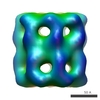
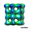
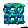
| Title | Structural basis of allosteric changes in the GroEL mutant Arg197-->Ala. | |||||||||
 Map data Map data | R197A Mutant of GroEL with 50uM ATp | |||||||||
 Sample Sample |
| |||||||||
| Biological species |  | |||||||||
| Method | single particle reconstruction / cryo EM / Resolution: 25.0 Å | |||||||||
 Authors Authors | White HE / Chen S / Roseman AM / Yifrach O / Horovitz A / SAibil HR | |||||||||
 Citation Citation | Journal: Nat Struct Biol / Year: 1997 Title: Structural basis of allosteric changes in the GroEL mutant Arg197-->Ala. | |||||||||
| History |
|
- Structure visualization
| Movie |
 Movie viewer Movie viewer |
|---|---|
| Structure viewer | EM map: SurfViewMolmilJmol/JSmol |
| Supplemental images |
 UCSF Chimera
- Downloads & links
-EMDB archive
| Map data | emd_1292.map.gz | 983.4 KB | EMDB map data format | |
|---|---|---|---|---|
| Header (meta data) | emd-1292-v30.xmlemd-1292.xml | 7.4 KB 7.4 KB | Display Display | EMDB header |
| Images |  1292.gif 1292.gif | 122.8 KB | ||
| Archive directory |  http://ftp.pdbj.org/pub/emdb/structures/EMD-1292ftp://ftp.pdbj.org/pub/emdb/structures/EMD-1292 http://ftp.pdbj.org/pub/emdb/structures/EMD-1292ftp://ftp.pdbj.org/pub/emdb/structures/EMD-1292 | HTTPS FTP |
-Related structure data


-Links
| EMDB pages | EMDB (EBI/PDBe) / EMDataResource |
|---|
-Map
| File | Download / File: emd_1292.map.gz / Format: CCP4 / Size: 1001 KB / Type: IMAGE STORED AS FLOATING POINT NUMBER (4 BYTES) | ||||||||||||||||||||||||||||||||||||||||||||||||||||||||||||||||||||
|---|---|---|---|---|---|---|---|---|---|---|---|---|---|---|---|---|---|---|---|---|---|---|---|---|---|---|---|---|---|---|---|---|---|---|---|---|---|---|---|---|---|---|---|---|---|---|---|---|---|---|---|---|---|---|---|---|---|---|---|---|---|---|---|---|---|---|---|---|---|
| Annotation | R197A Mutant of GroEL with 50uM ATp | ||||||||||||||||||||||||||||||||||||||||||||||||||||||||||||||||||||
| Projections & slices | Image control
Images are generated by Spider. | ||||||||||||||||||||||||||||||||||||||||||||||||||||||||||||||||||||
| Voxel size | X=Y=Z: 5.6 Å | ||||||||||||||||||||||||||||||||||||||||||||||||||||||||||||||||||||
| Density |
| ||||||||||||||||||||||||||||||||||||||||||||||||||||||||||||||||||||
| Symmetry | Space group: 1 | ||||||||||||||||||||||||||||||||||||||||||||||||||||||||||||||||||||
| Details | EMDB XML:
CCP4 map header:
| ||||||||||||||||||||||||||||||||||||||||||||||||||||||||||||||||||||
 Z (Sec.)
 Y (Row.)
 X (Col.)


-Supplemental data
- Sample components
-Entire : R197A mutant of GroEL, 50uM ATP
| Entire | Name: R197A mutant of GroEL, 50uM ATP |
|---|---|
| Components |
|
-Supramolecule #1000: R197A mutant of GroEL, 50uM ATP
| Supramolecule | Name: R197A mutant of GroEL, 50uM ATP / type: sample / ID: 1000 / Oligomeric state: 14-mer / Number unique components: 1 |
|---|
-Macromolecule #1: R197A mutant of GroEL
| Macromolecule | Name: R197A mutant of GroEL / type: protein_or_peptide / ID: 1 / Details: MUTANT / Number of copies: 1 / Oligomeric state: 14-mer / Recombinant expression: Yes |
|---|---|
| Source (natural) | Organism: |
-Experimental details
-Structure determination
| Method | cryo EM |
|---|---|
 Processing Processing | single particle reconstruction |
| Aggregation state | particle |
-Sample preparation
| Concentration | 1 mg/mL |
|---|---|
| Buffer | pH: 7.5 / Details: 20 mM Tris, 10mM KCl, 8mmM MgCl2,50uM ATP |
| Grid | Details: holey carbon |
| Vitrification | Cryogen name: ETHANE Timed resolved state: Vitrified within 5 secs of ATP addition |
- Electron microscopy
| Microscope | JEOL 1200EX |
|---|---|
| Image recording | Category: FILM / Film or detector model: AGFA SCIENTA FILM / Digitization - Scanner: OTHER / Digitization - Sampling interval: 10 µm |
| Electron beam | Acceleration voltage: 120 kV / Electron source: TUNGSTEN HAIRPIN |
| Electron optics | Illumination mode: OTHER / Imaging mode: OTHER |
| Sample stage | Specimen holder: Eucentric / Specimen holder model: OTHER |
-Image processing
| Final reconstruction | Applied symmetry - Point group: C7 (7 fold cyclic) / Algorithm: OTHER / Resolution.type: BY AUTHOR / Resolution: 25.0 Å / Software - Name: SPIDER |
|---|
-Atomic model buiding 1
| Details | Manual fitting in O of an apical domain of GroEL |
|---|---|
| Refinement | Protocol: RIGID BODY FIT |
