Model: Quantifoil R2/1 / Material: MOLYBDENUM / Mesh: 200 / Support film - Material: CARBON / Support film - topology: HOLEY
Vitrification
Cryogen name: ETHANE-PROPANE / Instrument: HOMEMADE PLUNGER Details: The grids were mounted on a manual plunger, blotted from the back side using Whatman paper #1 (Sigma-Aldrich) and plunged into a 2:1 ethane:propane mixture cooled down by liquid nitrogen by ...Details: The grids were mounted on a manual plunger, blotted from the back side using Whatman paper #1 (Sigma-Aldrich) and plunged into a 2:1 ethane:propane mixture cooled down by liquid nitrogen by the Martinsried-Plunger..
Details
A dissected intact Drosophila nervous system was deposited on an EM grid.
Cryo protectant
10% glycerol
Sectioning
Focused ion beam - Instrument: OTHER / Focused ion beam - Ion: OTHER / Focused ion beam - Voltage: 30 kV / Focused ion beam - Current: 0.05 nA / Focused ion beam - Duration: 3600 sec. / Focused ion beam - Temperature: 90 K / Focused ion beam - Initial thickness: 80000 nm / Focused ion beam - Final thickness: 140 nm Focused ion beam - Details: To prepare thin electron transparent lamellae into the tissue, plunge-frozen grids were first mounted into Autogrid frames (FEI). The grids were then mounted into a dual- ...Focused ion beam - Details: To prepare thin electron transparent lamellae into the tissue, plunge-frozen grids were first mounted into Autogrid frames (FEI). The grids were then mounted into a dual-beam Quanta 3D FIB/SEM (FEI) using a custom-built transfer shuttle and a cryo-transfer system (PP3000T, Quorum). The samples were kept at -180 C throughout FIB milling by the cryo-stage. To improve SEM imaging, a thin layer of pure metallic Pt was sputtered onto the sample under cryo conditions in the PP3000T transfer system to increase its electrical conductivity. The following parameters were used: 10 mA sputtering current, 500 V between stage and sputtering target and 30 s of exposure at 4.5x10⁻² mbar. To interpret and annotate the topographical anatomy of the tissue, overview maps of the EM grid were acquired by SEM at 10 kV at 100-250x magnification (object pixel size 1.1-0.4 um) and by secondary electrons induced by the Ga+ focused ion beam at 30 kV at 338x magnification (object pixel size 0.7 um). To protect the milling front of the lamellae, gaseous organic platinum was frozen on top of the grid using a gas injection system. To prevent bending of the lamella during the preparation, micro-expansion joints were milled left and right of the intended lamella preparation site. 10-20 um wide lamellae were prepared into the tissue with the ion beam at 30 kV at shallow angles (8-14 deg) in four consecutive steps: for the thicker tissue regions (e.g. VNC or skeletal muscle), the areas above and below the intended lamella were first removed with an ion beam current of 5 nA and 10 um spacing. This step was not necessary for thinner tissues such as the peripheral nerves. Further rectangular patterns were defined above and below the intended lamella with 2 um spacing for the rough milling step (ion beam current of 500-1000 pA), followed by fine milling with 800 nm spacing (100 pA) and a final polishing step down to the final lamella thickness of 100-200 nm (50 pA). To reach a uniform thickness, the lamella was tilted by +-0.5 deg and milled on each side separately with 50 pA current. The thickness of the lamella during the polishing step was assessed by SEM at 3-5 kV, 4.1 pA: the loss of charging effects in the lamella, visualized as the vanishing of bright areas, indicates a thickness <300 nm at 5 kV or <200 nm at 3 kV. Biological structures inside the lamella at the surface were imaged at each step by SEM at 2.5 kV, 4.1 pA in integration mode (64x). To reduce lamella charging during phase plate cryo-ET data acquisition, a thin layer of pure metallic Pt was sputtered onto the lamella under cryo conditions in the PP3000T transfer system with the following parameters: 5 mA sputtering current, 500 V between stage and sputtering target and 10 s of exposure at 4.5x10⁻² mbar.. The value given for _emd_sectioning_focused_ion_beam.instrument is FEI Quanta 3D FIB/SEM. This is not in a list of allowed values {'DB235', 'OTHER'} so OTHER is written into the XML file.
-
Electron microscopy
Microscope
FEI TITAN KRIOS
Specialist optics
Phase plate: VOLTA PHASE PLATE / Energy filter - Name: GIF Quantum LS / Energy filter - Slit width: 20 eV
Details
Additional Volta phase plate alignment.
Image recording
Film or detector model: GATAN K2 SUMMIT (4k x 4k) / Detector mode: COUNTING / Digitization - Dimensions - Width: 3868 pixel / Digitization - Dimensions - Height: 3868 pixel / Average electron dose: 3.0 e/Å2
Electron beam
Acceleration voltage: 300 kV / Electron source: FIELD EMISSION GUN
In the structure databanks used in Yorodumi, some data are registered as the other names, "COVID-19 virus" and "2019-nCoV". Here are the details of the virus and the list of structure data.
Jan 31, 2019. EMDB accession codes are about to change! (news from PDBe EMDB page)
EMDB accession codes are about to change! (news from PDBe EMDB page)
The allocation of 4 digits for EMDB accession codes will soon come to an end. Whilst these codes will remain in use, new EMDB accession codes will include an additional digit and will expand incrementally as the available range of codes is exhausted. The current 4-digit format prefixed with “EMD-” (i.e. EMD-XXXX) will advance to a 5-digit format (i.e. EMD-XXXXX), and so on. It is currently estimated that the 4-digit codes will be depleted around Spring 2019, at which point the 5-digit format will come into force.
The EM Navigator/Yorodumi systems omit the EMD- prefix.
Related info.:Q: What is EMD? / ID/Accession-code notation in Yorodumi/EM Navigator
Yorodumi is a browser for structure data from EMDB, PDB, SASBDB, etc.
This page is also the successor to EM Navigator detail page, and also detail information page/front-end page for Omokage search.
The word "yorodu" (or yorozu) is an old Japanese word meaning "ten thousand". "mi" (miru) is to see.
Related info.:EMDB / PDB / SASBDB / Comparison of 3 databanks / Yorodumi Search / Aug 31, 2016. New EM Navigator & Yorodumi / Yorodumi Papers / Jmol/JSmol / Function and homology information / Changes in new EM Navigator and Yorodumi
 Movie
Movie Controller

 Yorodumi
 Open data
 Basic information
 Map data
 Sample
 Authors
 Germany,
  China, 3 items
  Citation
 Structure visualization
 Movie viewer
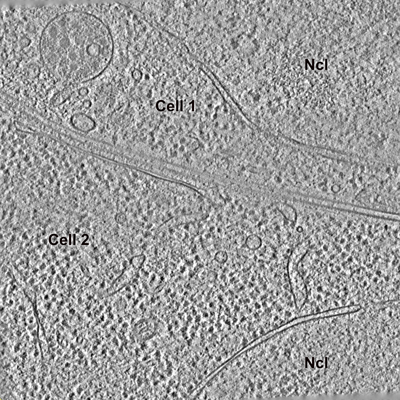
 Downloads & links
 emd_12727.png
 http://ftp.pdbj.org/pub/emdb/structures/EMD-12727

 Z (Sec.)
 Y (Row.)
 X (Col.)


 Sample components
 Processing
 Electron microscopy
 FIELD EMISSION GUN
