Journal: Front Mol Biosci / Year: 2021 Title: HEMNMA-3D: Cryo Electron Tomography Method Based on Normal Mode Analysis to Study Continuous Conformational Variability of Macromolecular Complexes. Authors: Mohamad Harastani / Mikhail Eltsov / Amélie Leforestier / Slavica Jonic / Abstract: Cryogenic electron tomography (cryo-ET) allows structural determination of biomolecules in their native environment (). Its potential of providing information on the dynamics of macromolecular ...Cryogenic electron tomography (cryo-ET) allows structural determination of biomolecules in their native environment (). Its potential of providing information on the dynamics of macromolecular complexes in cells is still largely unexploited, due to the challenges of the data analysis. The crowded cell environment and continuous conformational changes of complexes make difficult disentangling the data heterogeneity. We present HEMNMA-3D, which is, to the best of our knowledge, the first method for analyzing cryo electron subtomograms in terms of continuous conformational changes of complexes. HEMNMA-3D uses a combination of elastic and rigid-body 3D-to-3D iterative alignments of a flexible 3D reference (atomic structure or electron microscopy density map) to match the conformation, orientation, and position of the complex in each subtomogram. The elastic matching combines molecular mechanics simulation (Normal Mode Analysis of the 3D reference) and experimental, subtomogram data analysis. The rigid-body alignment includes compensation for the missing wedge, due to the limited tilt angle of cryo-ET. The conformational parameters (amplitudes of normal modes) of the complexes in subtomograms obtained through the alignment are processed to visualize the distribution of conformations in a space of lower dimension (typically, 2D or 3D) referred to as space of conformations. This allows a visually interpretable insight into the dynamics of the complexes, by calculating 3D averages of subtomograms with similar conformations from selected (densest) regions and by recording movies of the 3D reference's displacement along selected trajectories through the densest regions. We describe HEMNMA-3D and show its validation using synthetic datasets. We apply HEMNMA-3D to an experimental dataset describing nucleosome conformational variability. HEMNMA-3D software is available freely (open-source) as part of ContinuousFlex plugin of Scipion V3.0 (http://scipion.i2pc.es).
History
Deposition
Mar 31, 2021
-
Header (metadata) release
May 26, 2021
-
Map release
May 26, 2021
-
Update
May 26, 2021
-
Current status
May 26, 2021
Processing site: PDBe / Status: Released
-
Structure visualization
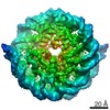
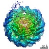
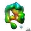
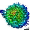
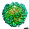
Movie
Surface view with section colored by density value
EMPIAR-10679 (Title: Subtomograms of nucleosomes extracted from cryo-tomograms of Drosophila melanogaster embryos Data size: 666.3 MB Data #1: Unaligned subtomograms of nucleosomes extracted from in situ cryo-tomograms of Drosophila melanogaster embryonic brain [subtomograms])
Model: C-flat-2/2 / Material: COPPER/PALLADIUM / Mesh: 200 / Support film - Material: CARBON
Vitrification
Cryogen name: OTHER / Details: high pressure freezer HPM010 (ABRA Fluid AG).
Details
Drosophila melanogaster embryos were high-pressure frozen. Vitreous sections were cut with a nominal thickness of 75 nm and collected onto 200 mesh C-flat grids
-
Electron microscopy
Microscope
FEI TITAN KRIOS
Image recording
Film or detector model: GATAN K2 SUMMIT (4k x 4k) / Detector mode: COUNTING / Average electron dose: 1.5 e/Å2
Electron beam
Acceleration voltage: 300 kV / Electron source: FIELD EMISSION GUN
Electron optics
Illumination mode: OTHER / Imaging mode: BRIGHT FIELD / Nominal defocus min: 3.5 µm / Nominal magnification: 64000
In the structure databanks used in Yorodumi, some data are registered as the other names, "COVID-19 virus" and "2019-nCoV". Here are the details of the virus and the list of structure data.
Jan 31, 2019. EMDB accession codes are about to change! (news from PDBe EMDB page)
EMDB accession codes are about to change! (news from PDBe EMDB page)
The allocation of 4 digits for EMDB accession codes will soon come to an end. Whilst these codes will remain in use, new EMDB accession codes will include an additional digit and will expand incrementally as the available range of codes is exhausted. The current 4-digit format prefixed with “EMD-” (i.e. EMD-XXXX) will advance to a 5-digit format (i.e. EMD-XXXXX), and so on. It is currently estimated that the 4-digit codes will be depleted around Spring 2019, at which point the 5-digit format will come into force.
The EM Navigator/Yorodumi systems omit the EMD- prefix.
Related info.:Q: What is EMD? / ID/Accession-code notation in Yorodumi/EM Navigator
Yorodumi is a browser for structure data from EMDB, PDB, SASBDB, etc.
This page is also the successor to EM Navigator detail page, and also detail information page/front-end page for Omokage search.
The word "yorodu" (or yorozu) is an old Japanese word meaning "ten thousand". "mi" (miru) is to see.
Related info.:EMDB / PDB / SASBDB / Comparison of 3 databanks / Yorodumi Search / Aug 31, 2016. New EM Navigator & Yorodumi / Yorodumi Papers / Jmol/JSmol / Function and homology information / Changes in new EM Navigator and Yorodumi
 Movie
Movie Controller

 Yorodumi
 Open data
 Basic information
 Map data
 Sample
 Function and homology information
 Authors
 France,
  Germany, 4 items
  Citation
 Structure visualization
 Downloads & links
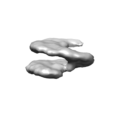 emd_12699.png
 http://ftp.pdbj.org/pub/emdb/structures/EMD-12699


 Z (Sec.)
 Y (Row.)
 X (Col.)


 Sample components
 Processing
 Electron microscopy
 FIELD EMISSION GUN
