 Movie
Movie Controller

[English] 日本語
 Yorodumi
- EMDB-1265: Structural changes of bacteriophage phi29 upon DNA packaging and ... -
+ Open data

- Basic information
| Entry | Database: EMDB / ID: EMD-1265 | |||||||||
|---|---|---|---|---|---|---|---|---|---|---|
| Title | Structural changes of bacteriophage phi29 upon DNA packaging and release. | |||||||||
 Map data Map data | Structure of bacteriophage phi29 mature head | |||||||||
 Sample Sample |
| |||||||||
| Biological species |   Bacillus phage phi29 (virus) Bacillus phage phi29 (virus) | |||||||||
| Method | single particle reconstruction / cryo EM / Resolution: 16.0 Å | |||||||||
 Authors Authors | Xiang Y / Morais MC / Battisti AJ / Grimes S / Jardine PJ / Anderson DL / Rossmann MG | |||||||||
 Citation Citation | Journal: EMBO J / Year: 2006 Title: Structural changes of bacteriophage phi29 upon DNA packaging and release. Authors: Ye Xiang / Marc C Morais / Anthony J Battisti / Shelley Grimes / Paul J Jardine / Dwight L Anderson / Michael G Rossmann /  Abstract: Cryo-electron microscopy three-dimensional reconstructions have been made of mature and of emptied bacteriophage phi29 particles without making symmetry assumptions. Comparisons of these structures ...Cryo-electron microscopy three-dimensional reconstructions have been made of mature and of emptied bacteriophage phi29 particles without making symmetry assumptions. Comparisons of these structures with each other and with the phi29 prohead indicate how conformational changes might initiate successive steps of assembly and infection. The 12 adsorption capable 'appendages' were found to have a structure homologous to the bacteriophage P22 tailspikes. Two of the appendages are extended radially outwards, away from the long axis of the virus, whereas the others are around and parallel to the phage axis. The appendage orientations are correlated with the symmetry-mismatched positions of the five-fold related head fibers, suggesting a mechanism for partial cell wall digestion upon rotation of the head about the tail when initiating infection. The narrow end of the head-tail connector is expanded in the mature virus. Gene product 3, bound to the 5' ends of the genome, appears to be positioned within the expanded connector, which may potentiate the release of DNA-packaging machine components, creating a binding site for attachment of the tail. | |||||||||
| History |
|
- Structure visualization
| Movie |
 Movie viewer Movie viewer |
|---|---|
| Structure viewer | EM map: SurfViewMolmilJmol/JSmol |
| Supplemental images |

- Downloads & links
-EMDB archive
| Map data | emd_1265.map.gz | 15.6 MB | EMDB map data format | |
|---|---|---|---|---|
| Header (meta data) | emd-1265-v30.xmlemd-1265.xml | 9.2 KB 9.2 KB | Display Display | EMDB header |
| Images | 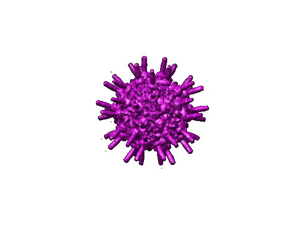 1265.gif 1265.gif | 20.6 KB | ||
| Archive directory |  http://ftp.pdbj.org/pub/emdb/structures/EMD-1265ftp://ftp.pdbj.org/pub/emdb/structures/EMD-1265 http://ftp.pdbj.org/pub/emdb/structures/EMD-1265ftp://ftp.pdbj.org/pub/emdb/structures/EMD-1265 | HTTPS FTP |
-Validation report
| Summary document | emd_1265_validation.pdf.gz | 268.1 KB | Display | EMDB validaton report |
|---|---|---|---|---|
| Full document | emd_1265_full_validation.pdf.gz | 267.2 KB | Display | |
| Data in XML | emd_1265_validation.xml.gz | 6.3 KB | Display | |
| Arichive directory | https://ftp.pdbj.org/pub/emdb/validation_reports/EMD-1265ftp://ftp.pdbj.org/pub/emdb/validation_reports/EMD-1265 | HTTPS FTP |
-Related structure data


-Links
| EMDB pages | EMDB (EBI/PDBe) / EMDataResource |
|---|
-Map
| File | Download / File: emd_1265.map.gz / Format: CCP4 / Size: 29.8 MB / Type: IMAGE STORED AS FLOATING POINT NUMBER (4 BYTES) | ||||||||||||||||||||||||||||||||||||||||||||||||||||||||||||||||||||
|---|---|---|---|---|---|---|---|---|---|---|---|---|---|---|---|---|---|---|---|---|---|---|---|---|---|---|---|---|---|---|---|---|---|---|---|---|---|---|---|---|---|---|---|---|---|---|---|---|---|---|---|---|---|---|---|---|---|---|---|---|---|---|---|---|---|---|---|---|---|
| Annotation | Structure of bacteriophage phi29 mature head | ||||||||||||||||||||||||||||||||||||||||||||||||||||||||||||||||||||
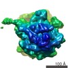
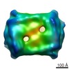
| Projections & slices | Image control
Images are generated by Spider. | ||||||||||||||||||||||||||||||||||||||||||||||||||||||||||||||||||||
| Voxel size | X=Y=Z: 4.24 Å | ||||||||||||||||||||||||||||||||||||||||||||||||||||||||||||||||||||
| Density |
| ||||||||||||||||||||||||||||||||||||||||||||||||||||||||||||||||||||
| Symmetry | Space group: 1 | ||||||||||||||||||||||||||||||||||||||||||||||||||||||||||||||||||||
| Details | EMDB XML:
CCP4 map header:
| ||||||||||||||||||||||||||||||||||||||||||||||||||||||||||||||||||||
 Z (Sec.)
 Y (Row.)
 X (Col.)


-Supplemental data
- Sample components
-Entire : phi29 mature particle
| Entire | Name: phi29 mature particle |
|---|---|
| Components |
|
-Supramolecule #1000: phi29 mature particle
| Supramolecule | Name: phi29 mature particle / type: sample / ID: 1000 Oligomeric state: capsid protein forms T3 Q5 prolate icosahedron Number unique components: 1 |
|---|---|
| Molecular weight | Theoretical: 33.6 MDa |
-Supramolecule #1: Bacillus phage phi29
| Supramolecule | Name: Bacillus phage phi29 / type: virus / ID: 1 / Name.synonym: M phi29, phi29 mature particle / NCBI-ID: 10756 / Sci species name: Bacillus phage phi29 / Virus type: VIRION / Virus isolate: SPECIES / Virus enveloped: Yes / Virus empty: No / Syn species name: M phi29, phi29 mature particle |
|---|---|
| Host (natural) | Organism:  |
| Molecular weight | Experimental: 33.6 MDa |
| Virus shell | Shell ID: 1 / Name: capsid-Length-T / Diameter: 530 Å / T number (triangulation number): 3 |
| Virus shell | Shell ID: 2 / Name: capsid-Width-Q / Diameter: 430 Å / T number (triangulation number): 5 |
-Experimental details
-Structure determination
| Method | cryo EM |
|---|---|
 Processing Processing | single particle reconstruction |
| Aggregation state | particle |
-Sample preparation
| Concentration | 1 mg/mL |
|---|---|
| Buffer | pH: 7.8 Details: 25 mM Tris-HCl pH7.8, 5mM MgCl2, 50mM NaCl and 2mM sodium azide |
| Grid | Details: holey carbon |
| Vitrification | Cryogen name: ETHANE |
- Electron microscopy
| Microscope | FEI/PHILIPS CM300FEG/T |
|---|---|
| Specialist optics | Energy filter - Name: FEI |
| Image recording | Category: FILM / Film or detector model: KODAK SO-163 FILM / Digitization - Scanner: ZEISS SCAI / Digitization - Sampling interval: 7 µm / Number real images: 68 / Average electron dose: 20 e/Å2 / Details: after scanning, images binned by a factor of 2 / Bits/pixel: 8 |
| Electron beam | Acceleration voltage: 300 kV / Electron source:  FIELD EMISSION GUN FIELD EMISSION GUN |
| Electron optics | Illumination mode: FLOOD BEAM / Imaging mode: BRIGHT FIELD / Cs: 2 mm / Nominal defocus max: 7.0 µm / Nominal defocus min: 2.0 µm / Nominal magnification: 33000 |
| Sample stage | Specimen holder: Side entry liquid nitrogen-cooled cryo specimen holder Specimen holder model: GATAN LIQUID NITROGEN |
-Image processing
| CTF correction | Details: phase flip and amplitude correction for each micrograph |
|---|---|
| Final reconstruction | Applied symmetry - Point group: C5 (5 fold cyclic) / Resolution.type: BY AUTHOR / Resolution: 16.0 Å / Resolution method: FSC 0.5 CUT-OFF / Software - Name: EMAN / Number images used: 12184 |
| Final two d classification | Number classes: 20 |
