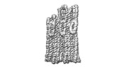
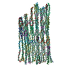
+ データを開く
- 基本情報
基本情報
| 登録情報 | データベース: EMDB / ID: EMD-12516 | |||||||||||||||
|---|---|---|---|---|---|---|---|---|---|---|---|---|---|---|---|---|
| タイトル | B-brick bare in 5 mM Mg2+ | |||||||||||||||
 マップデータ マップデータ | Composite map from Multibody and PostProcessing job | |||||||||||||||
 試料 試料 |
| |||||||||||||||
 キーワード キーワード | DNA Origami / DNA | |||||||||||||||
| 生物種 | synthetic construct (人工物) | |||||||||||||||
| 手法 | 単粒子再構成法 / クライオ電子顕微鏡法 / 解像度: 10.38 Å | |||||||||||||||
 データ登録者 データ登録者 | Bertosin E / Stoemmer P | |||||||||||||||
| 資金援助 |  ドイツ, 4件 ドイツ, 4件
| |||||||||||||||
 引用 引用 |  ジャーナル: ACS Nano / 年: 2021 ジャーナル: ACS Nano / 年: 2021タイトル: Cryo-Electron Microscopy and Mass Analysis of Oligolysine-Coated DNA Nanostructures. 著者: Eva Bertosin / Pierre Stömmer / Elija Feigl / Maximilian Wenig / Maximilian N Honemann / Hendrik Dietz /  要旨: Cationic coatings can enhance the stability of synthetic DNA objects in low ionic strength environments such as physiological fluids. Here, we used single-particle cryo-electron microscopy (cryo-EM), ...Cationic coatings can enhance the stability of synthetic DNA objects in low ionic strength environments such as physiological fluids. Here, we used single-particle cryo-electron microscopy (cryo-EM), pseudoatomic model fitting, and single-molecule mass photometry to study oligolysine and polyethylene glycol (PEG)-oligolysine-coated multilayer DNA origami objects. The coatings preserve coarse structural features well on a resolution of multiple nanometers but can also induce deformations such as twisting and bending. Higher-density coatings also led to internal structural deformations in the DNA origami test objects, in which a designed honeycomb-type helical lattice was deformed into a more square-lattice-like pattern. Under physiological ionic strength, where the uncoated objects disassembled, the coated objects remained intact but they shrunk in the helical direction and expanded in the direction perpendicular to the helical axis. Helical details like major/minor grooves and crossover locations were not discernible in cryo-EM maps that we determined of DNA origami coated with oligolysine and PEG-oligolysine, whereas these features were visible in cryo-EM maps determined from the uncoated reference objects. Blunt-ended double-helical interfaces remained accessible underneath the coating and may be used for the formation of multimeric DNA origami assemblies that rely on stacking interactions between blunt-ended helices. The ionic strength requirements for forming multimers from coated DNA origami differed from those needed for uncoated objects. Using single-molecule mass photometry, we found that the mass of coated DNA origami objects prior to and after incubation in low ionic strength physiological conditions remained unchanged. This finding indicated that the coating effectively prevented strand dissociation but also that the coating itself remained stable in place. Our results validate oligolysine coatings as a powerful stabilization method for DNA origami but also reveal several potential points of failure that experimenters should watch to avoid working with false premises. | |||||||||||||||
| 履歴 |
|
- 構造の表示
構造の表示
| ムービー |
 ムービービューア ムービービューア |
|---|---|
| 構造ビューア | EMマップ:  SurfView SurfView Molmil Molmil Jmol/JSmol Jmol/JSmol |
| 添付画像 |
- ダウンロードとリンク
-EMDBアーカイブ
| マップデータ |  emd_12516.map.gz emd_12516.map.gz | 7.9 MB |  EMDBマップデータ形式 EMDBマップデータ形式 | |
|---|---|---|---|---|
| ヘッダ (付随情報) |  emd-12516-v30.xml emd-12516-v30.xml emd-12516.xml emd-12516.xml | 93.4 KB 93.4 KB | 表示 表示 |  EMDBヘッダ EMDBヘッダ |
| FSC (解像度算出) |  emd_12516_fsc.xml emd_12516_fsc.xml | 10.8 KB | 表示 |  FSCデータファイル FSCデータファイル |
| 画像 |  emd_12516.png emd_12516.png | 56 KB | ||
| Filedesc metadata |  emd-12516.cif.gz emd-12516.cif.gz | 9.4 KB | ||
| その他 |  emd_12516_additional_1.map.gz emd_12516_additional_1.map.gz | 5.9 MB | ||
| アーカイブディレクトリ |  http://ftp.pdbj.org/pub/emdb/structures/EMD-12516 http://ftp.pdbj.org/pub/emdb/structures/EMD-12516 ftp://ftp.pdbj.org/pub/emdb/structures/EMD-12516 ftp://ftp.pdbj.org/pub/emdb/structures/EMD-12516 | HTTPS FTP |
-検証レポート
| 文書・要旨 |  emd_12516_validation.pdf.gz emd_12516_validation.pdf.gz | 432.1 KB | 表示 |  EMDB検証レポート EMDB検証レポート |
|---|---|---|---|---|
| 文書・詳細版 |  emd_12516_full_validation.pdf.gz emd_12516_full_validation.pdf.gz | 431.7 KB | 表示 | |
| XML形式データ |  emd_12516_validation.xml.gz emd_12516_validation.xml.gz | 12 KB | 表示 | |
| CIF形式データ |  emd_12516_validation.cif.gz emd_12516_validation.cif.gz | 15.8 KB | 表示 | |
| アーカイブディレクトリ |  https://ftp.pdbj.org/pub/emdb/validation_reports/EMD-12516 https://ftp.pdbj.org/pub/emdb/validation_reports/EMD-12516 ftp://ftp.pdbj.org/pub/emdb/validation_reports/EMD-12516 ftp://ftp.pdbj.org/pub/emdb/validation_reports/EMD-12516 | HTTPS FTP |
-関連構造データ
- リンク
リンク
| EMDBのページ |  EMDB (EBI/PDBe) / EMDB (EBI/PDBe) /  EMDataResource EMDataResource |
|---|---|
| 「今月の分子」の関連する項目 |
- マップ
マップ
| ファイル |  ダウンロード / ファイル: emd_12516.map.gz / 形式: CCP4 / 大きさ: 103 MB / タイプ: IMAGE STORED AS FLOATING POINT NUMBER (4 BYTES) ダウンロード / ファイル: emd_12516.map.gz / 形式: CCP4 / 大きさ: 103 MB / タイプ: IMAGE STORED AS FLOATING POINT NUMBER (4 BYTES) | ||||||||||||||||||||||||||||||||||||||||||||||||||||||||||||
|---|---|---|---|---|---|---|---|---|---|---|---|---|---|---|---|---|---|---|---|---|---|---|---|---|---|---|---|---|---|---|---|---|---|---|---|---|---|---|---|---|---|---|---|---|---|---|---|---|---|---|---|---|---|---|---|---|---|---|---|---|---|
| 注釈 | Composite map from Multibody and PostProcessing job | ||||||||||||||||||||||||||||||||||||||||||||||||||||||||||||
| 投影像・断面図 | 画像のコントロール
画像は Spider により作成 | ||||||||||||||||||||||||||||||||||||||||||||||||||||||||||||
| ボクセルのサイズ | X=Y=Z: 2.319 Å | ||||||||||||||||||||||||||||||||||||||||||||||||||||||||||||
| 密度 |
| ||||||||||||||||||||||||||||||||||||||||||||||||||||||||||||
| 対称性 | 空間群: 1 | ||||||||||||||||||||||||||||||||||||||||||||||||||||||||||||
| 詳細 | EMDB XML:
CCP4マップ ヘッダ情報:
| ||||||||||||||||||||||||||||||||||||||||||||||||||||||||||||
-添付データ
-追加マップ: Refined and post-processed map
| ファイル | emd_12516_additional_1.map | ||||||||||||
|---|---|---|---|---|---|---|---|---|---|---|---|---|---|
| 注釈 | Refined and post-processed map | ||||||||||||
| 投影像・断面図 |
| ||||||||||||
| 密度ヒストグラム |
- 試料の構成要素
+全体 : B-brick bare in 5 mM Mg2+
+超分子 #1: B-brick bare in 5 mM Mg2+
+分子 #1: SCAFFOLD STRAND
+分子 #2: STAPLE STRAND
+分子 #3: STAPLE STRAND
+分子 #4: STAPLE STRAND
+分子 #5: STAPLE STRAND
+分子 #6: STAPLE STRAND
+分子 #7: STAPLE STRAND
+分子 #8: STAPLE STRAND
+分子 #9: STAPLE STRAND
+分子 #10: STAPLE STRAND
+分子 #11: STAPLE STRAND
+分子 #12: STAPLE STRAND
+分子 #13: STAPLE STRAND
+分子 #14: STAPLE STRAND
+分子 #15: STAPLE STRAND
+分子 #16: STAPLE STRAND
+分子 #17: STAPLE STRAND
+分子 #18: STAPLE STRAND
+分子 #19: STAPLE STRAND
+分子 #20: STAPLE STRAND
+分子 #21: STAPLE STRAND
+分子 #22: STAPLE STRAND
+分子 #23: STAPLE STRAND
+分子 #24: STAPLE STRAND
+分子 #25: STAPLE STRAND
+分子 #26: STAPLE STRAND
+分子 #27: STAPLE STRAND
+分子 #28: STAPLE STRAND
+分子 #29: STAPLE STRAND
+分子 #30: STAPLE STRAND
+分子 #31: STAPLE STRAND
+分子 #32: STAPLE STRAND
+分子 #33: STAPLE STRAND
+分子 #34: STAPLE STRAND
+分子 #35: STAPLE STRAND
+分子 #36: STAPLE STRAND
+分子 #37: STAPLE STRAND
+分子 #38: STAPLE STRAND
+分子 #39: STAPLE STRAND
+分子 #40: STAPLE STRAND
+分子 #41: STAPLE STRAND
+分子 #42: STAPLE STRAND
+分子 #43: STAPLE STRAND
+分子 #44: STAPLE STRAND
+分子 #45: STAPLE STRAND
+分子 #46: STAPLE STRAND
+分子 #47: STAPLE STRAND
+分子 #48: STAPLE STRAND
+分子 #49: STAPLE STRAND
+分子 #50: STAPLE STRAND
+分子 #51: STAPLE STRAND
+分子 #52: STAPLE STRAND
+分子 #53: STAPLE STRAND
+分子 #54: STAPLE STRAND
+分子 #55: STAPLE STRAND
+分子 #56: STAPLE STRAND
+分子 #57: STAPLE STRAND
+分子 #58: STAPLE STRAND
+分子 #59: STAPLE STRAND
+分子 #60: STAPLE STRAND
+分子 #61: STAPLE STRAND
+分子 #62: STAPLE STRAND
+分子 #63: STAPLE STRAND
+分子 #64: STAPLE STRAND
+分子 #65: STAPLE STRAND
+分子 #66: STAPLE STRAND
+分子 #67: STAPLE STRAND
+分子 #68: STAPLE STRAND
+分子 #69: STAPLE STRAND
+分子 #70: STAPLE STRAND
+分子 #71: STAPLE STRAND
+分子 #72: STAPLE STRAND
+分子 #73: STAPLE STRAND
+分子 #74: STAPLE STRAND
+分子 #75: STAPLE STRAND
+分子 #76: STAPLE STRAND
+分子 #77: STAPLE STRAND
+分子 #78: STAPLE STRAND
+分子 #79: STAPLE STRAND
-実験情報
-構造解析
| 手法 | クライオ電子顕微鏡法 |
|---|---|
 解析 解析 | 単粒子再構成法 |
| 試料の集合状態 | particle |
- 試料調製
試料調製
| 緩衝液 | pH: 7.5 |
|---|---|
| 凍結 | 凍結剤: ETHANE |
- 電子顕微鏡法
| 顕微鏡 | FEI TITAN KRIOS |
|---|---|
| 撮影 | フィルム・検出器のモデル: FEI FALCON III (4k x 4k) 検出モード: INTEGRATING / 平均電子線量: 50.0 e/Å2 |
| 電子線 | 加速電圧: 300 kV / 電子線源:  FIELD EMISSION GUN FIELD EMISSION GUN |
| 電子光学系 | 照射モード: FLOOD BEAM / 撮影モード: BRIGHT FIELD |
| 実験機器 |  モデル: Titan Krios / 画像提供: FEI Company |
 ムービー
ムービー コントローラー


 Z (Sec.)
 Y (Row.)
 X (Col.)


