 Movie
Movie Controller

[English] 日本語
 Yorodumi
- EMDB-1218: The structural basis for regulated assembly and function of the t... -
+ Open data

- Basic information
| Entry | Database: EMDB / ID: EMD-1218 | |||||||||
|---|---|---|---|---|---|---|---|---|---|---|
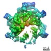
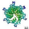
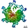
| Title | The structural basis for regulated assembly and function of the transcriptional activator NtrC. | |||||||||
 Map data Map data | 3D map of full-length, BeF-activated NtrC in the ADP-AlF state | |||||||||
 Sample Sample |
| |||||||||
| Biological species |  Salmonella enterica subsp. enterica serovar Typhimurium (bacteria) Salmonella enterica subsp. enterica serovar Typhimurium (bacteria) | |||||||||
| Method | single particle reconstruction / negative staining / Resolution: 28.0 Å | |||||||||
 Authors Authors | De Carlo S / Chen B / Hoover TR / Kondrashkina E / Nogales E / Nixon BT | |||||||||
 Citation Citation | Journal: Genes Dev / Year: 2006 Title: The structural basis for regulated assembly and function of the transcriptional activator NtrC. Authors: Sacha De Carlo / Baoyu Chen / Timothy R Hoover / Elena Kondrashkina / Eva Nogales / B Tracy Nixon /  Abstract: In two-component signal transduction, an input triggers phosphorylation of receiver domains that regulate the status of output modules. One such module is the AAA+ ATPase domain in bacterial enhancer- ...In two-component signal transduction, an input triggers phosphorylation of receiver domains that regulate the status of output modules. One such module is the AAA+ ATPase domain in bacterial enhancer-binding proteins that remodel the sigma(54) form of RNA polymerase. We report X-ray solution scattering and electron microscopy structures of the activated, full-length nitrogen-regulatory protein C (NtrC) showing a novel mechanism for regulation of AAA+ ATPase assembly via the juxtaposition of the receiver domains and ATPase ring. Accompanying the hydrolysis cycle that is required for transcriptional activation, we observed major order-disorder changes in the GAFTGA loops involved in sigma(54) binding, as well as in the DNA-binding domains. | |||||||||
| History |
|
- Structure visualization
| Movie |
 Movie viewer Movie viewer |
|---|---|
| Structure viewer | EM map: SurfViewMolmilJmol/JSmol |
| Supplemental images |
 UCSF Chimera
- Downloads & links
-EMDB archive
| Map data | emd_1218.map.gz | 5.2 MB | EMDB map data format | |
|---|---|---|---|---|
| Header (meta data) | emd-1218-v30.xmlemd-1218.xml | 9.1 KB 9.1 KB | Display Display | EMDB header |
| Images | 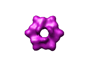 1218.gif 1218.gif | 9.5 KB | ||
| Archive directory |  http://ftp.pdbj.org/pub/emdb/structures/EMD-1218ftp://ftp.pdbj.org/pub/emdb/structures/EMD-1218 http://ftp.pdbj.org/pub/emdb/structures/EMD-1218ftp://ftp.pdbj.org/pub/emdb/structures/EMD-1218 | HTTPS FTP |
-Related structure data
| Similar structure data |
|---|
-Links
| EMDB pages | EMDB (EBI/PDBe) / EMDataResource |
|---|
-Map
| File | Download / File: emd_1218.map.gz / Format: CCP4 / Size: 7.3 MB / Type: IMAGE STORED AS FLOATING POINT NUMBER (4 BYTES) | ||||||||||||||||||||||||||||||||||||||||||||||||||||||||||||||||||||
|---|---|---|---|---|---|---|---|---|---|---|---|---|---|---|---|---|---|---|---|---|---|---|---|---|---|---|---|---|---|---|---|---|---|---|---|---|---|---|---|---|---|---|---|---|---|---|---|---|---|---|---|---|---|---|---|---|---|---|---|---|---|---|---|---|---|---|---|---|---|
| Annotation | 3D map of full-length, BeF-activated NtrC in the ADP-AlF state | ||||||||||||||||||||||||||||||||||||||||||||||||||||||||||||||||||||
| Projections & slices | Image control
Images are generated by Spider. | ||||||||||||||||||||||||||||||||||||||||||||||||||||||||||||||||||||
| Voxel size | X=Y=Z: 2.56 Å | ||||||||||||||||||||||||||||||||||||||||||||||||||||||||||||||||||||
| Density |
| ||||||||||||||||||||||||||||||||||||||||||||||||||||||||||||||||||||
| Symmetry | Space group: 1 | ||||||||||||||||||||||||||||||||||||||||||||||||||||||||||||||||||||
| Details | EMDB XML:
CCP4 map header:
| ||||||||||||||||||||||||||||||||||||||||||||||||||||||||||||||||||||
 Z (Sec.)
 Y (Row.)
 X (Col.)


-Supplemental data
- Sample components
-Entire : Full length, active NtrC
| Entire | Name: Full length, active NtrC |
|---|---|
| Components |
|
-Supramolecule #1000: Full length, active NtrC
| Supramolecule | Name: Full length, active NtrC / type: sample / ID: 1000 / Details: Activated with Mg/BeF / Oligomeric state: Hexamer / Number unique components: 1 |
|---|---|
| Molecular weight | Experimental: 300 KDa / Theoretical: 300 KDa / Method: Sedimentation Velocity, Analytical centrifugation |
-Macromolecule #1: Transcription activator
| Macromolecule | Name: Transcription activator / type: protein_or_peptide / ID: 1 / Name.synonym: Enhancer-binding protein Details: Baers following mutations: S160F, R456A, N457A, R461A Number of copies: 1 / Oligomeric state: Hexamer / Recombinant expression: Yes |
|---|---|
| Source (natural) | Organism: Salmonella enterica subsp. enterica serovar Typhimurium (bacteria) synonym: Bacteria / Cell: Salmonella typhimurium |
| Molecular weight | Experimental: 300 KDa / Theoretical: 300 KDa |
| Recombinant expression | Organism: |
-Experimental details
-Structure determination
| Method | negative staining |
|---|---|
 Processing Processing | single particle reconstruction |
| Aggregation state | particle |
-Sample preparation
| Concentration | 0.05 mg/mL |
|---|---|
| Buffer | pH: 8.2 Details: 200mM KCl, 20mM Tris, 1mM nucleotide, 0.2 mM BeCl2 and 5mM NaF, 5mM MgCl2, 5% Trehalose |
| Staining | Type: NEGATIVE / Details: 3% uranyl acetate |
| Vitrification | Cryogen name: NONE |
- Electron microscopy
| Microscope | FEI TECNAI 12 |
|---|---|
| Alignment procedure | Legacy - Astigmatism: corrected / Legacy - Electron beam tilt params: 0 |
| Image recording | Category: FILM / Film or detector model: KODAK SO-163 FILM / Digitization - Scanner: OTHER / Digitization - Sampling interval: 6.7 µm / Number real images: 18 / Average electron dose: 20 e/Å2 / Bits/pixel: 14 |
| Tilt angle min | 0 |
| Electron beam | Acceleration voltage: 120 kV / Electron source: LAB6 |
| Electron optics | Calibrated magnification: 49767 / Illumination mode: FLOOD BEAM / Imaging mode: BRIGHT FIELD / Cs: 6.2 mm / Nominal defocus max: 1.5 µm / Nominal defocus min: 0.9 µm / Nominal magnification: 50000 |
| Sample stage | Specimen holder: simple tilt / Specimen holder model: OTHER / Tilt angle max: 50 |
-Image processing
| Final reconstruction | Applied symmetry - Point group: C3 (3 fold cyclic) / Algorithm: OTHER / Resolution.type: BY AUTHOR / Resolution: 28.0 Å / Resolution method: FSC 0.5 CUT-OFF / Software - Name: SPIDER / Number images used: 3500 |
|---|
