[English] 日本語
 Yorodumi
- EMDB-12162: Microtubule doublet structure from WT Chlamydomonas as a control ... -
+ Open data
- Basic information
| Entry | Database: EMDB / ID: EMD-12162 | |||||||||
|---|---|---|---|---|---|---|---|---|---|---|
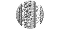
| Title | Microtubule doublet structure from WT Chlamydomonas as a control for MOT7 mutants | |||||||||
 Map data Map data | Cilia from WT Chlamydomonas as a control data for mutants, EMD-12160 and EMD-12161 | |||||||||
 Sample Sample |
| |||||||||
| Biological species |  | |||||||||
| Method | subtomogram averaging / cryo EM / Resolution: 25.0 Å | |||||||||
 Authors Authors | Obbineni JM / Kutomi O / Yamamoto R / Nakagiri Y / Imai H / Noga A / Zimmermann N / Ishikawa T / Wakabayashi K / Kon T / Inaba K | |||||||||
| Funding support |  Switzerland, Switzerland,  Japan, 2 items Japan, 2 items
| |||||||||
 Citation Citation |  Journal: Sci Adv / Year: 2021 Journal: Sci Adv / Year: 2021Title: A dynein-associated photoreceptor protein prevents ciliary acclimation to blue light. Authors: Osamu Kutomi / Ryosuke Yamamoto / Keiko Hirose / Katsutoshi Mizuno / Yuuhei Nakagiri / Hiroshi Imai / Akira Noga / Jagan Mohan Obbineni / Noemi Zimmermann / Masako Nakajima / Daisuke Shibata ...Authors: Osamu Kutomi / Ryosuke Yamamoto / Keiko Hirose / Katsutoshi Mizuno / Yuuhei Nakagiri / Hiroshi Imai / Akira Noga / Jagan Mohan Obbineni / Noemi Zimmermann / Masako Nakajima / Daisuke Shibata / Misa Shibata / Kogiku Shiba / Masaki Kita / Hideo Kigoshi / Yui Tanaka / Yuya Yamasaki / Yuma Asahina / Chihong Song / Mami Nomura / Mamoru Nomura / Ayako Nakajima / Mia Nakachi / Lixy Yamada / Shiori Nakazawa / Hitoshi Sawada / Kazuyoshi Murata / Kaoru Mitsuoka / Takashi Ishikawa / Ken-Ichi Wakabayashi / Takahide Kon / Kazuo Inaba /    Abstract: Light-responsive regulation of ciliary motility is known to be conducted through modulation of dyneins, but the mechanism is not fully understood. Here, we report a novel subunit of the two-headed ...Light-responsive regulation of ciliary motility is known to be conducted through modulation of dyneins, but the mechanism is not fully understood. Here, we report a novel subunit of the two-headed f/I1 inner arm dynein, named DYBLUP, in animal spermatozoa and a unicellular green alga. This subunit contains a BLUF (sensors of blue light using FAD) domain that appears to directly modulate dynein activity in response to light. DYBLUP (dynein-associated BLUF protein) mediates the connection between the f/I1 motor domain and the tether complex that links the motor to the doublet microtubule. lacking the DYBLUP ortholog shows both positive and negative phototaxis but becomes acclimated and attracted to high-intensity blue light. These results suggest a mechanism to avoid toxic strong light via direct photoregulation of dyneins. | |||||||||
| History |
|
- Structure visualization
| Movie |
 Movie viewer Movie viewer |
|---|---|
| Structure viewer | EM map:  SurfView SurfView Molmil Molmil Jmol/JSmol Jmol/JSmol |
| Supplemental images |
- Downloads & links
-EMDB archive
| Map data |  emd_12162.map.gz emd_12162.map.gz | 8.3 MB |  EMDB map data format EMDB map data format | |
|---|---|---|---|---|
| Header (meta data) |  emd-12162-v30.xml emd-12162-v30.xml emd-12162.xml emd-12162.xml | 12.8 KB 12.8 KB | Display Display |  EMDB header EMDB header |
| Images |  emd_12162.png emd_12162.png | 100.6 KB | ||
| Archive directory |  http://ftp.pdbj.org/pub/emdb/structures/EMD-12162 http://ftp.pdbj.org/pub/emdb/structures/EMD-12162 ftp://ftp.pdbj.org/pub/emdb/structures/EMD-12162 ftp://ftp.pdbj.org/pub/emdb/structures/EMD-12162 | HTTPS FTP |
-Validation report
| Summary document |  emd_12162_validation.pdf.gz emd_12162_validation.pdf.gz | 238.9 KB | Display |  EMDB validaton report EMDB validaton report |
|---|---|---|---|---|
| Full document |  emd_12162_full_validation.pdf.gz emd_12162_full_validation.pdf.gz | 238 KB | Display | |
| Data in XML |  emd_12162_validation.xml.gz emd_12162_validation.xml.gz | 5.6 KB | Display | |
| Arichive directory |  https://ftp.pdbj.org/pub/emdb/validation_reports/EMD-12162 https://ftp.pdbj.org/pub/emdb/validation_reports/EMD-12162 ftp://ftp.pdbj.org/pub/emdb/validation_reports/EMD-12162 ftp://ftp.pdbj.org/pub/emdb/validation_reports/EMD-12162 | HTTPS FTP |
-Related structure data
| Related structure data | C: citing same article ( |
|---|---|
| Similar structure data |
- Links
Links
| EMDB pages |  EMDB (EBI/PDBe) / EMDB (EBI/PDBe) /  EMDataResource EMDataResource |
|---|
- Map
Map
| File |  Download / File: emd_12162.map.gz / Format: CCP4 / Size: 16.8 MB / Type: IMAGE STORED AS FLOATING POINT NUMBER (4 BYTES) Download / File: emd_12162.map.gz / Format: CCP4 / Size: 16.8 MB / Type: IMAGE STORED AS FLOATING POINT NUMBER (4 BYTES) | ||||||||||||||||||||||||||||||||||||||||||||||||||||||||||||
|---|---|---|---|---|---|---|---|---|---|---|---|---|---|---|---|---|---|---|---|---|---|---|---|---|---|---|---|---|---|---|---|---|---|---|---|---|---|---|---|---|---|---|---|---|---|---|---|---|---|---|---|---|---|---|---|---|---|---|---|---|---|
| Annotation | Cilia from WT Chlamydomonas as a control data for mutants, EMD-12160 and EMD-12161 | ||||||||||||||||||||||||||||||||||||||||||||||||||||||||||||
| Projections & slices | Image control
Images are generated by Spider. | ||||||||||||||||||||||||||||||||||||||||||||||||||||||||||||
| Voxel size | X=Y=Z: 8.4 Å | ||||||||||||||||||||||||||||||||||||||||||||||||||||||||||||
| Density |
| ||||||||||||||||||||||||||||||||||||||||||||||||||||||||||||
| Symmetry | Space group: 1 | ||||||||||||||||||||||||||||||||||||||||||||||||||||||||||||
| Details | EMDB XML:
CCP4 map header:
| ||||||||||||||||||||||||||||||||||||||||||||||||||||||||||||
-Supplemental data
- Sample components
-Entire : doublet microtubules from cilia
| Entire | Name: doublet microtubules from cilia |
|---|---|
| Components |
|
-Supramolecule #1: doublet microtubules from cilia
| Supramolecule | Name: doublet microtubules from cilia / type: organelle_or_cellular_component / ID: 1 / Parent: 0 Details: Average of subtomograms extracted from microtubule doublet parts of cilia |
|---|---|
| Source (natural) | Organism:  |
-Experimental details
-Structure determination
| Method | cryo EM |
|---|---|
 Processing Processing | subtomogram averaging |
| Aggregation state | filament |
- Sample preparation
| Buffer | pH: 7 / Details: TAP medium |
|---|---|
| Grid | Model: Quantifoil R3.5/1 / Material: COPPER / Mesh: 300 / Support film - Material: CARBON / Support film - topology: HOLEY ARRAY / Pretreatment - Type: PLASMA CLEANING |
| Vitrification | Cryogen name: ETHANE / Chamber humidity: 90 % / Instrument: GATAN CRYOPLUNGE 3 |
| Details | cilia isolated from the cell and demembraned |
- Electron microscopy
| Microscope | FEI TITAN KRIOS |
|---|---|
| Specialist optics | Energy filter - Name: GIF Quantum LS / Energy filter - Slit width: 25 eV |
| Image recording | Film or detector model: GATAN K2 SUMMIT (4k x 4k) / Detector mode: COUNTING / Number grids imaged: 3 / Average exposure time: 1.0 sec. / Average electron dose: 60.0 e/Å2 |
| Electron beam | Acceleration voltage: 300 kV / Electron source:  FIELD EMISSION GUN FIELD EMISSION GUN |
| Electron optics | C2 aperture diameter: 100.0 µm / Illumination mode: FLOOD BEAM / Imaging mode: BRIGHT FIELD / Cs: 2.7 mm / Nominal defocus max: 5.0 µm / Nominal defocus min: 4.0 µm / Nominal magnification: 30000 |
| Sample stage | Specimen holder model: FEI TITAN KRIOS AUTOGRID HOLDER / Cooling holder cryogen: NITROGEN |
| Experimental equipment |  Model: Titan Krios / Image courtesy: FEI Company |
- Image processing
| Final reconstruction | Applied symmetry - Point group: C1 (asymmetric) / Resolution.type: BY AUTHOR / Resolution: 25.0 Å / Resolution method: FSC 0.5 CUT-OFF / Software - Name:  IMOD / Number subtomograms used: 3000 IMOD / Number subtomograms used: 3000 |
|---|---|
| Extraction | Number tomograms: 20 / Number images used: 4000 |
| CTF correction | Software - Name:  IMOD IMOD |
| Final angle assignment | Type: PROJECTION MATCHING |
 Movie
Movie Controller


 Z (Sec.)
 Y (Row.)
 X (Col.)


