 Movie
Movie Controller

[English] 日本語
 Yorodumi
- EMDB-12146: Brevibacterium linens encapsulin-associated Dye-decolorizing pero... -
+ Open data

- Basic information
| Entry |  | |||||||||
|---|---|---|---|---|---|---|---|---|---|---|
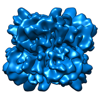
| Title | Brevibacterium linens encapsulin-associated Dye-decolorizing peroxidase | |||||||||
 Map data Map data | Brevibacterium linens encapsulin-associated Dye-decolorizing peroxidase | |||||||||
 Sample Sample |
| |||||||||
| Biological species |  Brevibacterium linens (bacteria) Brevibacterium linens (bacteria) | |||||||||
| Method | single particle reconstruction / cryo EM / Resolution: 8.2 Å | |||||||||
 Authors Authors | Allende-Ballestero C / Luque D / Klem R / Cornelissen JJLM / Caston JR | |||||||||
 Citation Citation | Journal: To Be Published Title: Three-dimensional cryoEM structure of Brevibacterium linens encapsulin Authors: Allende-Ballestero C / Luque D / Klem R / Cornelissen JJLM / Caston JR | |||||||||
| History |
|
- Structure visualization
| Supplemental images |
|---|
- Downloads & links
-EMDB archive
| Map data | emd_12146.map.gz | 12 MB |  EMDB map data format EMDB map data format | |
|---|---|---|---|---|
| Header (meta data) | emd-12146-v30.xmlemd-12146.xml | 12.6 KB 12.6 KB | Display Display | EMDB header |
| Images |  emd_12146.png emd_12146.png | 223.6 KB | ||
| Archive directory |  http://ftp.pdbj.org/pub/emdb/structures/EMD-12146ftp://ftp.pdbj.org/pub/emdb/structures/EMD-12146 http://ftp.pdbj.org/pub/emdb/structures/EMD-12146ftp://ftp.pdbj.org/pub/emdb/structures/EMD-12146 | HTTPS FTP |
-Related structure data
-Links
| EMDB pages | EMDB (EBI/PDBe) / EMDataResource |
|---|
-Map
| File | Download / File: emd_12146.map.gz / Format: CCP4 / Size: 12.9 MB / Type: IMAGE STORED AS FLOATING POINT NUMBER (4 BYTES) | ||||||||||||||||||||||||||||||||||||
|---|---|---|---|---|---|---|---|---|---|---|---|---|---|---|---|---|---|---|---|---|---|---|---|---|---|---|---|---|---|---|---|---|---|---|---|---|---|
| Annotation | Brevibacterium linens encapsulin-associated Dye-decolorizing peroxidase | ||||||||||||||||||||||||||||||||||||
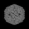
| Projections & slices | Image control
Images are generated by Spider. | ||||||||||||||||||||||||||||||||||||
| Voxel size | X=Y=Z: 1.047 Å | ||||||||||||||||||||||||||||||||||||
| Density |
| ||||||||||||||||||||||||||||||||||||
| Symmetry | Space group: 1 | ||||||||||||||||||||||||||||||||||||
| Details | EMDB XML:
|
 Z (Sec.)
 Y (Row.)
 X (Col.)


-Supplemental data
- Sample components
-Entire : Dye-decolorizing peroxidase
| Entire | Name: Dye-decolorizing peroxidase |
|---|---|
| Components |
|
-Supramolecule #1: Dye-decolorizing peroxidase
| Supramolecule | Name: Dye-decolorizing peroxidase / type: complex / ID: 1 / Parent: 0 / Macromolecule list: all |
|---|---|
| Source (natural) | Organism: Brevibacterium linens (bacteria) |
| Recombinant expression | Organism: |
| Molecular weight | Theoretical: 240 KDa |
-Macromolecule #1: Dye-decolorizing peroxidase
| Macromolecule | Name: Dye-decolorizing peroxidase / type: protein_or_peptide / ID: 1 / Enantiomer: LEVO |
|---|---|
| Source (natural) | Organism: Brevibacterium linens (bacteria) |
| Recombinant expression | Organism: |
| Sequence | String: MALPNGKTPQ HVLGPPAPAA VFLVLTVRSG AEAEAKDFLG DIAGVVRSVG FRAREDHLSC VTGIGAELWD RMFDAPRPAG LHPFIEQRGD VHTAPSTPGD LLFHIRARRM DLCFELARQL VGELGDAVSV VDEVHGFRYF DERDIMGFVD GTENPEDQEA VDSVFTPTGG ...String: MALPNGKTPQ HVLGPPAPAA VFLVLTVRSG AEAEAKDFLG DIAGVVRSVG FRAREDHLSC VTGIGAELWD RMFDAPRPAG LHPFIEQRGD VHTAPSTPGD LLFHIRARRM DLCFELARQL VGELGDAVSV VDEVHGFRYF DERDIMGFVD GTENPEDQEA VDSVFTPTGG DDPASSTYVI VQKYTHDMAA WEALSVEDQE AAFGRHKLSD MEFPDEDKAP NSHLILNTIE DEDGTEHKIV RDNMVFGSVE SGEFGTYFIG YAADVSVTEQ MLENMFIGNP RGTYDRILDF STAQTGGLFF VPSQDFLDDP DGELAAAEPS DAQNDDPASA SARVEETDPP NPASADDPAP ADDSLGIGSL RRRDQ |
-Experimental details
-Structure determination
| Method | cryo EM |
|---|---|
 Processing Processing | single particle reconstruction |
| Aggregation state | particle |
-Sample preparation
| Buffer | pH: 7.5 Component:
| ||||||
|---|---|---|---|---|---|---|---|
| Grid | Model: Quantifoil R2/2 / Material: COPPER/RHODIUM / Mesh: 400 / Support film - Material: CARBON / Support film - topology: HOLEY / Pretreatment - Type: GLOW DISCHARGE | ||||||
| Vitrification | Cryogen name: ETHANE / Chamber humidity: 95 % / Chamber temperature: 295 K / Instrument: FEI VITROBOT MARK IV |
- Electron microscopy
| Microscope | FEI TITAN KRIOS |
|---|---|
| Image recording | Film or detector model: GATAN K2 SUMMIT (4k x 4k) / Detector mode: COUNTING / Average electron dose: 38.85 e/Å2 |
| Electron beam | Acceleration voltage: 300 kV / Electron source:  FIELD EMISSION GUN FIELD EMISSION GUN |
| Electron optics | Calibrated defocus max: 3.2 µm / Calibrated defocus min: 1.0 µm / Illumination mode: FLOOD BEAM / Imaging mode: BRIGHT FIELD / Cs: 2.7 mm / Nominal defocus max: 3.2 µm / Nominal defocus min: 1.0 µm |
| Sample stage | Specimen holder model: FEI TITAN KRIOS AUTOGRID HOLDER / Cooling holder cryogen: NITROGEN |
| Experimental equipment |  Model: Titan Krios / Image courtesy: FEI Company |
+Image processing
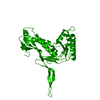
-Atomic model buiding 1
| Initial model | PDB ID: Chain - Chain ID: A |
|---|---|
| Refinement | Protocol: FLEXIBLE FIT |

