 Movie
Movie Controller

[English] 日本語
 Yorodumi
- EMDB-12093: Higher-order structures of the foot-and-mouth disease virus RNA-d... -
+ Open data

- Basic information
| Entry | Database: EMDB / ID: EMD-12093 | ||||||||||||||||||||||||
|---|---|---|---|---|---|---|---|---|---|---|---|---|---|---|---|---|---|---|---|---|---|---|---|---|---|
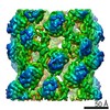
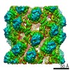
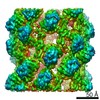
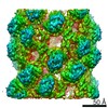
| Title | Higher-order structures of the foot-and-mouth disease virus RNA-dependent RNA polymerase required for dynamic inter-molecular interactions involved in viral genome replication | ||||||||||||||||||||||||
 Map data Map data | Helical reconstruction of fibril formed of foot and mouth disease virus RNA dependent RNA polymerase 3Dpol | ||||||||||||||||||||||||
 Sample Sample |
| ||||||||||||||||||||||||
| Function / homology |  Function and homology information Function and homology informationsymbiont-mediated perturbation of host chromatin organization / T=pseudo3 icosahedral viral capsid / host cell cytoplasmic vesicle membrane / ribonucleoside triphosphate phosphatase activity / channel activity / monoatomic ion transmembrane transport / clathrin-dependent endocytosis of virus by host cell / RNA helicase activity / viral protein processing / host cell endoplasmic reticulum membrane ...symbiont-mediated perturbation of host chromatin organization / T=pseudo3 icosahedral viral capsid / host cell cytoplasmic vesicle membrane / ribonucleoside triphosphate phosphatase activity / channel activity / monoatomic ion transmembrane transport / clathrin-dependent endocytosis of virus by host cell / RNA helicase activity / viral protein processing / host cell endoplasmic reticulum membrane / cysteine-type endopeptidase activity / viral RNA genome replication / RNA-directed RNA polymerase activity / virion attachment to host cell / DNA-templated transcription / host cell nucleus / structural molecule activity / proteolysis / RNA binding / ATP binding / metal ion binding Similarity search - Function | ||||||||||||||||||||||||
| Biological species |   Foot-and-mouth disease virus Foot-and-mouth disease virus | ||||||||||||||||||||||||
| Method | helical reconstruction / cryo EM / Resolution: 7.3 Å | ||||||||||||||||||||||||
 Authors Authors | Loundras EA / Streetley J / Herod MJ / Thompson R / Harris M / Bhella D / Stonehouse NJ | ||||||||||||||||||||||||
| Funding support |  United Kingdom, 7 items United Kingdom, 7 items
| ||||||||||||||||||||||||
 Citation Citation | Journal: Commun Biol / Year: 2022 Title: Higher-order structures of the foot-and-mouth disease virus RNA-dependent RNA polymerase required for genome replication. Authors: Eleni-Anna Loundras / James Streetley / Morgan R Herod / Rebecca Thompson / Mark Harris / David Bhella / Nicola J Stonehouse / Abstract: Replication of many positive-sense RNA viruses occurs within intracellular membrane-associated compartments. These are thought to provide a favourable environment for replication to occur, ...Replication of many positive-sense RNA viruses occurs within intracellular membrane-associated compartments. These are thought to provide a favourable environment for replication to occur, concentrating essential viral structural and nonstructural components, as well as protecting these components from host-cell pathogen recognition and innate immune responses. However, the details of the molecular interactions and dynamics within these structures is very limited. One of the key components of the replication machinery is the RNA-dependent RNA polymerase, RdRp. This enzyme has been shown to form higher-order fibrils in vitro. Here, using the RdRp from foot-and-mouth disease virus (termed 3D), we report fibril structures, solved at ~7-9 Å resolution by cryo-EM, revealing multiple conformations of a flexible assembly. Fitting high-resolution coordinates led to the definition of potential intermolecular interactions. We employed mutagenesis using a sub-genomic replicon system to probe the importance of these interactions for replication. We use these data to propose models for the role of higher-order 3D complexes as a dynamic scaffold within which RNA replication can occur. | ||||||||||||||||||||||||
| History |
|
- Structure visualization
| Movie |
Movie viewer |
|---|---|
| Structure viewer | EM map: SurfViewMolmilJmol/JSmol |
| Supplemental images |

- Downloads & links
-EMDB archive
| Map data | emd_12093.map.gz | 5.9 MB | EMDB map data format | |
|---|---|---|---|---|
| Header (meta data) | emd-12093-v30.xmlemd-12093.xml | 14.1 KB 14.1 KB | Display Display | EMDB header |
| FSC (resolution estimation) | emd_12093_fsc.xml | 7.2 KB | Display | FSC data file |
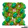
| Images | 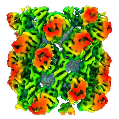 emd_12093.png emd_12093.png | 205 KB | ||
| Archive directory |  http://ftp.pdbj.org/pub/emdb/structures/EMD-12093ftp://ftp.pdbj.org/pub/emdb/structures/EMD-12093 http://ftp.pdbj.org/pub/emdb/structures/EMD-12093ftp://ftp.pdbj.org/pub/emdb/structures/EMD-12093 | HTTPS FTP |
-Related structure data
| Related structure data | C: citing same article ( |
|---|---|
| Similar structure data | |
| EM raw data | EMPIAR-10602 (Title: Higher-order structures of the foot-and-mouth disease virus RNA-dependent RNA polymerase required for dynamic inter-molecular interactions involved in viral genome replication Data size: 13.1 TB Data #1: Unaligned multi-frame micrographs of foot and mouth disease virus 3Dpol fibrils [micrographs - multiframe]) |

-Links
| EMDB pages | EMDB (EBI/PDBe) / EMDataResource |
|---|---|
| Related items in Molecule of the Month |


-Map
| File | Download / File: emd_12093.map.gz / Format: CCP4 / Size: 30.5 MB / Type: IMAGE STORED AS FLOATING POINT NUMBER (4 BYTES) | ||||||||||||||||||||||||||||||||||||||||||||||||||||||||||||
|---|---|---|---|---|---|---|---|---|---|---|---|---|---|---|---|---|---|---|---|---|---|---|---|---|---|---|---|---|---|---|---|---|---|---|---|---|---|---|---|---|---|---|---|---|---|---|---|---|---|---|---|---|---|---|---|---|---|---|---|---|---|
| Annotation | Helical reconstruction of fibril formed of foot and mouth disease virus RNA dependent RNA polymerase 3Dpol | ||||||||||||||||||||||||||||||||||||||||||||||||||||||||||||
| Projections & slices | Image control
Images are generated by Spider. | ||||||||||||||||||||||||||||||||||||||||||||||||||||||||||||
| Voxel size | X=Y=Z: 2.13 Å | ||||||||||||||||||||||||||||||||||||||||||||||||||||||||||||
| Density |
| ||||||||||||||||||||||||||||||||||||||||||||||||||||||||||||
| Symmetry | Space group: 1 | ||||||||||||||||||||||||||||||||||||||||||||||||||||||||||||
| Details | EMDB XML:
CCP4 map header:
| ||||||||||||||||||||||||||||||||||||||||||||||||||||||||||||
 Z (Sec.)
 Y (Row.)
 X (Col.)


-Supplemental data
- Sample components
-Entire : Helical fibril of foot and mouth disease virus RNA dependent RNA ...
| Entire | Name: Helical fibril of foot and mouth disease virus RNA dependent RNA polymerase (3Dpol) |
|---|---|
| Components |
|
-Supramolecule #1: Helical fibril of foot and mouth disease virus RNA dependent RNA ...
| Supramolecule | Name: Helical fibril of foot and mouth disease virus RNA dependent RNA polymerase (3Dpol) type: complex / ID: 1 / Parent: 0 |
|---|---|
| Source (natural) | Organism: Foot-and-mouth disease virus |
| Recombinant expression | Organism:  |
-Experimental details
-Structure determination
| Method | cryo EM |
|---|---|
 Processing Processing | helical reconstruction |
| Aggregation state | filament |
-Sample preparation
| Buffer | pH: 7 |
|---|---|
| Grid | Model: C-flat-1.2/1.3 / Material: COPPER / Mesh: 400 / Support film - Material: CARBON / Support film - topology: CONTINUOUS |
| Vitrification | Cryogen name: ETHANE / Chamber humidity: 100 % / Chamber temperature: 277 K / Instrument: FEI VITROBOT MARK IV Details: Specimen was loaded onto C-flat (1.2/1.3), 400 mesh holey carbon support films bearing a continuous carbon layer. Allowed to adsorb for one minute before blotting for 6-seconds and plunge freezing.. |
| Details | Fibrils were formed by mixing FMDV 3DPol with a 13mer of poly-adenosine RNA, oligo-d(T) primer and UTP in the presence of 5mM glutaraldehyde. |
- Electron microscopy
| Microscope | FEI TITAN KRIOS |
|---|---|
| Image recording | Film or detector model: FEI FALCON III (4k x 4k) / Detector mode: INTEGRATING / Number grids imaged: 2 / Number real images: 5417 / Average exposure time: 2.0 sec. / Average electron dose: 108.0 e/Å2 Details: The dose rate is 60 e/pix/s, giving a dose of 54 e/a2/second. We are taking a 2 second exposure, with 79 frames in total, giving 1.4 electrons/frame |
| Electron beam | Acceleration voltage: 300 kV / Electron source:  FIELD EMISSION GUN FIELD EMISSION GUN |
| Electron optics | Illumination mode: FLOOD BEAM / Imaging mode: BRIGHT FIELD / Cs: 2.7 mm / Nominal defocus max: 2.8000000000000003 µm / Nominal defocus min: 1.5 µm / Nominal magnification: 75000 |
| Sample stage | Specimen holder model: FEI TITAN KRIOS AUTOGRID HOLDER / Cooling holder cryogen: NITROGEN |
| Experimental equipment |  Model: Titan Krios / Image courtesy: FEI Company |
+Image processing

-Atomic model buiding 1
| Initial model | PDB ID: |
|---|---|
| Refinement | Protocol: RIGID BODY FIT |

