 Movie
Movie Controller

[English] 日本語
 Yorodumi
- EMDB-1188: An archaeal peptidase assembles into two different quaternary str... -
+ Open data

- Basic information
| Entry | Database: EMDB / ID: EMD-1188 | |||||||||
|---|---|---|---|---|---|---|---|---|---|---|
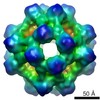
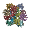
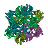
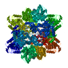
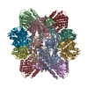
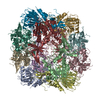
| Title | An archaeal peptidase assembles into two different quaternary structures: A tetrahedron and a giant octahedron. | |||||||||
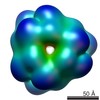
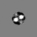
 Map data Map data | 3D volume of the tetrahedral aminopeptidase TET1 from Pyrococus horikoshii | |||||||||
 Sample Sample |
| |||||||||
| Biological species |   Pyrococcus horikoshii (archaea) Pyrococcus horikoshii (archaea) | |||||||||
| Method | single particle reconstruction / cryo EM / negative staining / Resolution: 14.0 Å | |||||||||
 Authors Authors | Schoehn G / Vellieux FM / Asuncion Dura M / Receveur-Brechot V / Fabry CM / Ruigrok RW / Ebel C / Roussel A / Franzetti B | |||||||||
 Citation Citation | Journal: J Biol Chem / Year: 2006 Title: An archaeal peptidase assembles into two different quaternary structures: A tetrahedron and a giant octahedron. Authors: Guy Schoehn / Frédéric M D Vellieux / M Asunción Durá / Véronique Receveur-Bréchot / Céline M S Fabry / Rob W H Ruigrok / Christine Ebel / Alain Roussel / Bruno Franzetti /  Abstract: Cellular proteolysis involves large oligomeric peptidases that play key roles in the regulation of many cellular processes. The cobalt-activated peptidase TET1 from the hyperthermophilic Archaea ...Cellular proteolysis involves large oligomeric peptidases that play key roles in the regulation of many cellular processes. The cobalt-activated peptidase TET1 from the hyperthermophilic Archaea Pyrococcus horikoshii (PhTET1) was found to assemble as a 12-subunit tetrahedron and as a 24-subunit octahedral particle. Both quaternary structures were solved by combining x-ray crystallography and cryoelectron microscopy data. The internal organization of the PhTET1 particles reveals highly self-compartmentalized systems made of networks of access channels extended by vast catalytic chambers. The two edifices display aminopeptidase activity, and their organizations indicate substrate navigation mechanisms different from those described in other large peptidase complexes. Compared with the tetrahedron, the octahedron forms a more expanded hollow structure, representing a new type of giant peptidase complex. PhTET1 assembles into two different quaternary structures because of quasi-equivalent contacts that previously have only been identified in viral capsids. | |||||||||
| History |
|
- Structure visualization
| Movie |
 Movie viewer Movie viewer |
|---|---|
| Structure viewer | EM map: SurfViewMolmilJmol/JSmol |
| Supplemental images |
 UCSF Chimera
- Downloads & links
-EMDB archive
| Map data | emd_1188.map.gz | 7.4 MB | EMDB map data format | |
|---|---|---|---|---|
| Header (meta data) | emd-1188-v30.xmlemd-1188.xml | 10.3 KB 10.3 KB | Display Display | EMDB header |
| Images |  1188.gif 1188.gif | 8.3 KB | ||
| Archive directory |  http://ftp.pdbj.org/pub/emdb/structures/EMD-1188ftp://ftp.pdbj.org/pub/emdb/structures/EMD-1188 http://ftp.pdbj.org/pub/emdb/structures/EMD-1188ftp://ftp.pdbj.org/pub/emdb/structures/EMD-1188 | HTTPS FTP |
-Validation report
| Summary document | emd_1188_validation.pdf.gz | 200.3 KB | Display | EMDB validaton report |
|---|---|---|---|---|
| Full document | emd_1188_full_validation.pdf.gz | 199.4 KB | Display | |
| Data in XML | emd_1188_validation.xml.gz | 5.4 KB | Display | |
| Arichive directory | https://ftp.pdbj.org/pub/emdb/validation_reports/EMD-1188ftp://ftp.pdbj.org/pub/emdb/validation_reports/EMD-1188 | HTTPS FTP |
-Related structure data

-Links
| EMDB pages | EMDB (EBI/PDBe) / EMDataResource |
|---|
-Map
| File | Download / File: emd_1188.map.gz / Format: CCP4 / Size: 7.8 MB / Type: IMAGE STORED AS FLOATING POINT NUMBER (4 BYTES) | ||||||||||||||||||||||||||||||||||||||||||||||||||||||||||||||||||||
|---|---|---|---|---|---|---|---|---|---|---|---|---|---|---|---|---|---|---|---|---|---|---|---|---|---|---|---|---|---|---|---|---|---|---|---|---|---|---|---|---|---|---|---|---|---|---|---|---|---|---|---|---|---|---|---|---|---|---|---|---|---|---|---|---|---|---|---|---|---|
| Annotation | 3D volume of the tetrahedral aminopeptidase TET1 from Pyrococus horikoshii | ||||||||||||||||||||||||||||||||||||||||||||||||||||||||||||||||||||
| Projections & slices | Image control
Images are generated by Spider. | ||||||||||||||||||||||||||||||||||||||||||||||||||||||||||||||||||||
| Voxel size | X=Y=Z: 2.8 Å | ||||||||||||||||||||||||||||||||||||||||||||||||||||||||||||||||||||
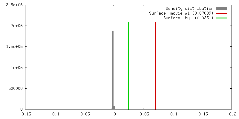
| Density |
| ||||||||||||||||||||||||||||||||||||||||||||||||||||||||||||||||||||
| Symmetry | Space group: 1 | ||||||||||||||||||||||||||||||||||||||||||||||||||||||||||||||||||||
| Details | EMDB XML:
CCP4 map header:
| ||||||||||||||||||||||||||||||||||||||||||||||||||||||||||||||||||||
 Z (Sec.)
 Y (Row.)
 X (Col.)


-Supplemental data
- Sample components
-Entire : TET1 metallopeptidase from Pyrococcus horikoshii
| Entire | Name: TET1 metallopeptidase from Pyrococcus horikoshii |
|---|---|
| Components |
|
-Supramolecule #1000: TET1 metallopeptidase from Pyrococcus horikoshii
| Supramolecule | Name: TET1 metallopeptidase from Pyrococcus horikoshii / type: sample / ID: 1000 / Oligomeric state: 12 mer / Number unique components: 1 |
|---|---|
| Molecular weight | Experimental: 400 KDa / Theoretical: 400 KDa / Method: Analytical ultracentrifugation |
-Macromolecule #1: cobalt activated metallopeptidase TET1
| Macromolecule | Name: cobalt activated metallopeptidase TET1 / type: protein_or_peptide / ID: 1 / Details: Pyrococcus horikoshii / Number of copies: 12 / Oligomeric state: 12-mer / Recombinant expression: Yes |
|---|---|
| Source (natural) | Organism: Pyrococcus horikoshii (archaea) / synonym: PH0519 / Tissue: Pyrococcus horikoshii / Cell: Pyrococcus horikoshii / Organelle: Pyrococcus horikoshii |
| Recombinant expression | Organism: Pyrococcus horikoshii (archaea) |
-Experimental details
-Structure determination
| Method | negative staining, cryo EM |
|---|---|
 Processing Processing | single particle reconstruction |
| Aggregation state | particle |
-Sample preparation
| Concentration | 1 mg/mL |
|---|---|
| Buffer | pH: 7.4 |
| Staining | Type: NEGATIVE Details: Quantifoil R2 1 grids (Quantifoil Micro Tools GmbH, Germany) were loaded with 4 ul of sample at 1 mg ml, blotted and rapidly frozen in liquid ethane within a liquid nitrogen bath using a Zeiss cryoplunger |
| Grid | Details: Quantifoil R2/1 grids |
| Vitrification | Cryogen name: ETHANE / Chamber temperature: 100 K / Instrument: OTHER / Details: Vitrification instrument: Zeiss cryoplunger |
- Electron microscopy
| Microscope | FEI/PHILIPS CM200T |
|---|---|
| Temperature | Min: 100 K / Average: 100 K |
| Alignment procedure | Legacy - Astigmatism: bjective lens astigmatism was corrected at |
| Date | Jan 1, 2002 |
| Image recording | Category: FILM / Film or detector model: KODAK SO-163 FILM / Digitization - Scanner: ZEISS SCAI / Digitization - Sampling interval: 14 µm / Number real images: 15 / Average electron dose: 15 e/Å2 / Od range: 1.2 / Bits/pixel: 8 |
| Electron beam | Acceleration voltage: 200 kV / Electron source: LAB6 |
| Electron optics | Illumination mode: SPOT SCAN / Imaging mode: BRIGHT FIELD / Cs: 2 mm / Nominal defocus max: 2.5 µm / Nominal defocus min: 1.0 µm / Nominal magnification: 50000 |
| Sample stage | Specimen holder: Eucentric / Specimen holder model: GATAN LIQUID NITROGEN |
-Image processing
| CTF correction | Details: ctfmix |
|---|---|
| Final reconstruction | Applied symmetry - Point group: T (tetrahedral) / Algorithm: OTHER / Resolution.type: BY AUTHOR / Resolution: 14.0 Å / Resolution method: OTHER / Software - Name: spider / Details: 8000 particles in 159 class average / Number images used: 8000 |
| Final two d classification | Number classes: 159 |
-Atomic model buiding 1
| Details | Protocol: Rigid Body. colores from Situs |
|---|---|
| Refinement | Protocol: RIGID BODY FIT |
