 Movie
Movie Controller

[English] 日本語
 Yorodumi
- EMDB-1154: Kelp fly virus: a novel group of insect picorna-like viruses as d... -
+ Open data

- Basic information
| Entry | Database: EMDB / ID: EMD-1154 | |||||||||
|---|---|---|---|---|---|---|---|---|---|---|
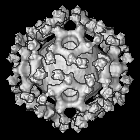
| Title | Kelp fly virus: a novel group of insect picorna-like viruses as defined by genome sequence analysis and a distinctive virion structure. | |||||||||
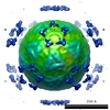
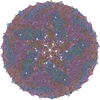
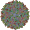
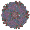
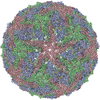
 Map data Map data | This is a complete icosahedral reconstruction of Kelp fly virus | |||||||||
 Sample Sample |
| |||||||||
| Biological species |  Kelp fly virus Kelp fly virus | |||||||||
| Method | single particle reconstruction / cryo EM / Resolution: 15.0 Å | |||||||||
 Authors Authors | Hartley CJ / Greenwood DR / Gilbert RJC | |||||||||
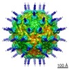
 Citation Citation | Journal: J Virol / Year: 2005 Title: Kelp fly virus: a novel group of insect picorna-like viruses as defined by genome sequence analysis and a distinctive virion structure. Authors: C J Hartley / D R Greenwood / R J C Gilbert / A Masoumi / K H J Gordon / T N Hanzlik / E E Fry / D I Stuart / P D Scotti /  Abstract: The complete genomic sequence of kelp fly virus (KFV), originally isolated from the kelp fly, Chaetocoelopa sydneyensis, has been determined. Analyses of its genomic and structural organization and ...The complete genomic sequence of kelp fly virus (KFV), originally isolated from the kelp fly, Chaetocoelopa sydneyensis, has been determined. Analyses of its genomic and structural organization and phylogeny show that it belongs to a hitherto undescribed group within the picorna-like virus superfamily. The single-stranded genomic RNA of KFV is 11,035 nucleotides in length and contains a single large open reading frame encoding a polypeptide of 3,436 amino acids with 5' and 3' untranslated regions of 384 and 343 nucleotides, respectively. The predicted amino acid sequence of the polypeptide shows that it has three regions. The N-terminal region contains sequences homologous to the baculoviral inhibitor of apoptosis repeat domain, an inhibitor of apoptosis commonly found in animals and in viruses with double-stranded DNA genomes. The second region contains at least two capsid proteins. The third region has three sequence motifs characteristic of replicase proteins of many plant and animal viruses, including a helicase, a 3C chymotrypsin-like protease, and an RNA-dependent RNA polymerase. Phylogenetic analysis of the replicase motifs shows that KFV forms a distinct and distant taxon within the picorna-like virus superfamily. Cryoelectron microscopy and image reconstruction of KFV to a resolution of 15 A reveals an icosahedral structure, with each of its 12 fivefold vertices forming a turret from the otherwise smooth surface of the 20-A-thick capsid. The architecture of the KFV capsid is unique among the members of the picornavirus superfamily for which structures have previously been determined. | |||||||||
| History |
|
- Structure visualization
| Movie |
 Movie viewer Movie viewer |
|---|---|
| Structure viewer | EM map: SurfViewMolmilJmol/JSmol |
| Supplemental images |
- Downloads & links
-EMDB archive
| Map data | emd_1154.map.gz | 1.1 MB | EMDB map data format | |
|---|---|---|---|---|
| Header (meta data) | emd-1154-v30.xmlemd-1154.xml | 9.8 KB 9.8 KB | Display Display | EMDB header |
| Images |  1154.gif 1154.gif | 68.5 KB | ||
| Archive directory |  http://ftp.pdbj.org/pub/emdb/structures/EMD-1154ftp://ftp.pdbj.org/pub/emdb/structures/EMD-1154 http://ftp.pdbj.org/pub/emdb/structures/EMD-1154ftp://ftp.pdbj.org/pub/emdb/structures/EMD-1154 | HTTPS FTP |
-Related structure data

-Links
| EMDB pages | EMDB (EBI/PDBe) / EMDataResource |
|---|
-Map
| File | Download / File: emd_1154.map.gz / Format: CCP4 / Size: 10.2 MB / Type: IMAGE STORED AS FLOATING POINT NUMBER (4 BYTES) | ||||||||||||||||||||||||||||||||||||||||||||||||||||||||||||||||||||
|---|---|---|---|---|---|---|---|---|---|---|---|---|---|---|---|---|---|---|---|---|---|---|---|---|---|---|---|---|---|---|---|---|---|---|---|---|---|---|---|---|---|---|---|---|---|---|---|---|---|---|---|---|---|---|---|---|---|---|---|---|---|---|---|---|---|---|---|---|---|
| Annotation | This is a complete icosahedral reconstruction of Kelp fly virus | ||||||||||||||||||||||||||||||||||||||||||||||||||||||||||||||||||||
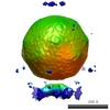
| Projections & slices | Image control
Images are generated by Spider. | ||||||||||||||||||||||||||||||||||||||||||||||||||||||||||||||||||||
| Voxel size | X=Y=Z: 4.38 Å | ||||||||||||||||||||||||||||||||||||||||||||||||||||||||||||||||||||
| Density |
| ||||||||||||||||||||||||||||||||||||||||||||||||||||||||||||||||||||
| Symmetry | Space group: 1 | ||||||||||||||||||||||||||||||||||||||||||||||||||||||||||||||||||||
| Details | EMDB XML:
CCP4 map header:
| ||||||||||||||||||||||||||||||||||||||||||||||||||||||||||||||||||||
 Z (Sec.)
 X (Row.)
 Y (Col.)


-Supplemental data
- Sample components
-Entire : Kelp fly virus
| Entire | Name: Kelp fly virus |
|---|---|
| Components |
|
-Supramolecule #1000: Kelp fly virus
| Supramolecule | Name: Kelp fly virus / type: sample / ID: 1000 Details: The sample displays evidence of a substoichiometric level of turret decoration. Oligomeric state: One icosahedral virion / Number unique components: 1 |
|---|---|
| Molecular weight | Experimental: 4.38 MDa Method: Volumetrically from the reconstruction and by comparison with the known volume to mass ratio of other insect viruses |
-Supramolecule #1: Kelp fly virus
| Supramolecule | Name: Kelp fly virus / type: virus / ID: 1 / Name.synonym: KFV / NCBI-ID: 340921 / Sci species name: Kelp fly virus / Virus type: VIRION / Virus isolate: SPECIES / Virus enveloped: No / Virus empty: No / Syn species name: KFV |
|---|---|
| Host (natural) | Organism:  Chaetocoelopa sydneyensis (fry) / synonym: INVERTEBRATES Chaetocoelopa sydneyensis (fry) / synonym: INVERTEBRATES |
| Molecular weight | Theoretical: 4.38 MDa |
| Virus shell | Shell ID: 1 / Name: Capsid / Diameter: 330 Å |
| Virus shell | Shell ID: 2 / Name: Capsid and turrets / Diameter: 450 Å |
-Experimental details
-Structure determination
| Method | cryo EM |
|---|---|
 Processing Processing | single particle reconstruction |
| Aggregation state | particle |
-Sample preparation
| Concentration | 1.0 mg/mL |
|---|---|
| Buffer | pH: 7.2 / Details: 100 mM TRIS, pH 7.2 |
| Grid | Details: 300 mesh copper grid, carbon lacey film |
| Vitrification | Cryogen name: ETHANE Method: Blot until grid pulls away from Whatman number 1 paper before plunging |
- Electron microscopy
| Microscope | FEI/PHILIPS CM200FEG |
|---|---|
| Temperature | Average: 100 K |
| Alignment procedure | Legacy - Astigmatism: objective lens astigmatism was corrected at |
| Date | Jun 1, 2001 |
| Image recording | Category: FILM / Film or detector model: KODAK SO-163 FILM / Digitization - Scanner: OTHER / Digitization - Sampling interval: 4.38 µm / Number real images: 27 / Average electron dose: 2 e/Å2 / Bits/pixel: 8 |
| Electron beam | Acceleration voltage: 200 kV / Electron source:  FIELD EMISSION GUN FIELD EMISSION GUN |
| Electron optics | Illumination mode: SPOT SCAN / Imaging mode: BRIGHT FIELD / Cs: 2 mm / Nominal defocus max: 7.0 µm / Nominal defocus min: 1.0 µm / Nominal magnification: 38000 |
| Sample stage | Specimen holder: Eucentric / Specimen holder model: GATAN LIQUID NITROGEN |
-Image processing
| CTF correction | Details: By micrograph |
|---|---|
| Final reconstruction | Applied symmetry - Point group: I (icosahedral) / Algorithm: OTHER / Resolution.type: BY AUTHOR / Resolution: 15.0 Å / Resolution method: FSC 0.5 CUT-OFF / Software - Name: SPIDER and GAP Details: Final map was calculated from CTF-corrected reconstruction with unique vertex presented a turret (EMD3965) by applying icosahedral symmetry to an icosahedral asymmetric unit using GAP Number images used: 3890 |
| Final angle assignment | Details: SPIDER |
