 Movie
Movie Controller

[English] 日本語
 Yorodumi
- EMDB-11416: Tomogram of GFP-a-synuclein inclusion in primary mouse neuron exp... -
+ Open data

- Basic information
| Entry | Database: EMDB / ID: EMD-11416 | |||||||||
|---|---|---|---|---|---|---|---|---|---|---|
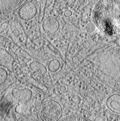
| Title | Tomogram of GFP-a-synuclein inclusion in primary mouse neuron expressing GFP-a-synuclein, seeded with MSA aggregates | |||||||||
 Map data Map data | Tomogram of GFP-a-synuclein inclusion in primary mouse neurons seeded with MSA aggregates | |||||||||
 Sample Sample |
| |||||||||
| Biological species |  | |||||||||
| Method | electron tomography / cryo EM | |||||||||
 Authors Authors | Trinkaus VA / Hartl FU / Fernandez-Busnadiego R | |||||||||
| Funding support |  Germany, 2 items Germany, 2 items
| |||||||||
 Citation Citation | Journal: Nat Commun / Year: 2021 Title: In situ architecture of neuronal α-Synuclein inclusions. Authors: Victoria A Trinkaus / Irene Riera-Tur / Antonio Martínez-Sánchez / Felix J B Bäuerlein / Qiang Guo / Thomas Arzberger / Wolfgang Baumeister / Irina Dudanova / Mark S Hipp / F Ulrich Hartl ...Authors: Victoria A Trinkaus / Irene Riera-Tur / Antonio Martínez-Sánchez / Felix J B Bäuerlein / Qiang Guo / Thomas Arzberger / Wolfgang Baumeister / Irina Dudanova / Mark S Hipp / F Ulrich Hartl / Rubén Fernández-Busnadiego /    Abstract: The molecular architecture of α-Synuclein (α-Syn) inclusions, pathognomonic of various neurodegenerative disorders, remains unclear. α-Syn inclusions were long thought to consist mainly of α-Syn ...The molecular architecture of α-Synuclein (α-Syn) inclusions, pathognomonic of various neurodegenerative disorders, remains unclear. α-Syn inclusions were long thought to consist mainly of α-Syn fibrils, but recent reports pointed to intracellular membranes as the major inclusion component. Here, we use cryo-electron tomography (cryo-ET) to image neuronal α-Syn inclusions in situ at molecular resolution. We show that inclusions seeded by α-Syn aggregates produced recombinantly or purified from patient brain consist of α-Syn fibrils crisscrossing a variety of cellular organelles. Using gold-labeled seeds, we find that aggregate seeding is predominantly mediated by small α-Syn fibrils, from which cytoplasmic fibrils grow unidirectionally. Detailed analysis of membrane interactions revealed that α-Syn fibrils do not contact membranes directly, and that α-Syn does not drive membrane clustering. Altogether, we conclusively demonstrate that neuronal α-Syn inclusions consist of α-Syn fibrils intermixed with membranous organelles, and illuminate the mechanism of aggregate seeding and cellular interaction. | |||||||||
| History |
|
- Structure visualization
| Movie |
 Movie viewer Movie viewer |
|---|---|
| Supplemental images |
- Downloads & links
-EMDB archive
| Map data | emd_11416.map.gz | 719 MB | EMDB map data format | |
|---|---|---|---|---|
| Header (meta data) | emd-11416-v30.xmlemd-11416.xml | 11.8 KB 11.8 KB | Display Display | EMDB header |
| Images |  emd_11416.png emd_11416.png | 348.1 KB | ||
| Archive directory |  http://ftp.pdbj.org/pub/emdb/structures/EMD-11416ftp://ftp.pdbj.org/pub/emdb/structures/EMD-11416 http://ftp.pdbj.org/pub/emdb/structures/EMD-11416ftp://ftp.pdbj.org/pub/emdb/structures/EMD-11416 | HTTPS FTP |
-Related structure data
-Links
| EMDB pages | EMDB (EBI/PDBe) / EMDataResource |
|---|
-Map
| File | Download / File: emd_11416.map.gz / Format: CCP4 / Size: 778.6 MB / Type: IMAGE STORED AS FLOATING POINT NUMBER (4 BYTES) | ||||||||||||||||||||||||||||||||||||||||||||||||||||||||||||
|---|---|---|---|---|---|---|---|---|---|---|---|---|---|---|---|---|---|---|---|---|---|---|---|---|---|---|---|---|---|---|---|---|---|---|---|---|---|---|---|---|---|---|---|---|---|---|---|---|---|---|---|---|---|---|---|---|---|---|---|---|---|
| Annotation | Tomogram of GFP-a-synuclein inclusion in primary mouse neurons seeded with MSA aggregates | ||||||||||||||||||||||||||||||||||||||||||||||||||||||||||||
| Projections & slices | Image control
Images are generated by Spider. generated in cubic-lattice coordinate | ||||||||||||||||||||||||||||||||||||||||||||||||||||||||||||
| Voxel size | X=Y=Z: 14.08 Å | ||||||||||||||||||||||||||||||||||||||||||||||||||||||||||||
| Density |
| ||||||||||||||||||||||||||||||||||||||||||||||||||||||||||||
| Symmetry | Space group: 1 | ||||||||||||||||||||||||||||||||||||||||||||||||||||||||||||
| Details | EMDB XML:
CCP4 map header:
| ||||||||||||||||||||||||||||||||||||||||||||||||||||||||||||
 Z (Sec.)
 Y (Row.)
 X (Col.)


-Supplemental data
- Sample components
-Entire : Tomogram of GFP-a-synuclein inclusion in primary mouse neuron exp...
| Entire | Name: Tomogram of GFP-a-synuclein inclusion in primary mouse neuron expressing GFP-a-synuclein, seeded with MSA aggregates |
|---|---|
| Components |
|
-Supramolecule #1: Tomogram of GFP-a-synuclein inclusion in primary mouse neuron exp...
| Supramolecule | Name: Tomogram of GFP-a-synuclein inclusion in primary mouse neuron expressing GFP-a-synuclein, seeded with MSA aggregates type: cell / ID: 1 / Parent: 0 / Macromolecule list: #1 |
|---|---|
| Source (natural) | Organism: |
-Experimental details
-Structure determination
| Method | cryo EM |
|---|---|
 Processing Processing | electron tomography |
| Aggregation state | cell |
-Sample preparation
| Buffer | pH: 7.4 |
|---|---|
| Grid | Model: Quantifoil R2/4 / Material: GOLD / Support film - Material: CARBON / Support film - topology: HOLEY / Pretreatment - Type: PLASMA CLEANING |
| Vitrification | Cryogen name: ETHANE-PROPANE / Chamber humidity: 80 % / Chamber temperature: 310 K / Instrument: FEI VITROBOT MARK IV |
| Cryo protectant | 10 % Glycerol |
| Sectioning | Focused ion beam - Instrument: OTHER / Focused ion beam - Ion: OTHER / Focused ion beam - Voltage: 30 kV / Focused ion beam - Current: 0.1 nA / Focused ion beam - Duration: 2400 sec. / Focused ion beam - Temperature: 77 K / Focused ion beam - Initial thickness: 1000 nm / Focused ion beam - Final thickness: 150 nm Focused ion beam - Details: The value given for _emd_sectioning_focused_ion_beam.instrument is FEI Quanta FIB. This is not in a list of allowed values {'DB235', 'OTHER'} so OTHER is written into the XML file. |
- Electron microscopy
| Microscope | FEI TITAN KRIOS |
|---|---|
| Temperature | Min: 70.0 K / Max: 75.0 K |
| Specialist optics | Energy filter - Name: GIF Bioquantum / Energy filter - Slit width: 20 eV |
| Image recording | Film or detector model: GATAN K2 SUMMIT (4k x 4k) / Detector mode: COUNTING / Digitization - Dimensions - Width: 3838 pixel / Digitization - Dimensions - Height: 3710 pixel / Digitization - Frames/image: 14-20 / Number grids imaged: 1 / Number real images: 50 / Average exposure time: 4.0 sec. / Average electron dose: 2.0 e/Å2 |
| Electron beam | Acceleration voltage: 300 kV / Electron source:  FIELD EMISSION GUN FIELD EMISSION GUN |
| Electron optics | C2 aperture diameter: 50.0 µm / Illumination mode: OTHER / Imaging mode: BRIGHT FIELD / Nominal defocus max: 7.0 µm / Nominal defocus min: 6.0 µm / Nominal magnification: 42000 |
| Sample stage | Specimen holder model: FEI TITAN KRIOS AUTOGRID HOLDER / Cooling holder cryogen: NITROGEN |
| Experimental equipment |  Model: Titan Krios / Image courtesy: FEI Company |
-Image processing
| Final reconstruction | Software - Name: IMOD (ver. 4.9.0) / Number images used: 50 |
|---|
