[English] 日本語
Yorodumi
- EMDB-11239: Map of Torpedo nicotinic acetylcholine receptor obtained from hel... -

+
Open data


ID or keywords:

Loading...

-
Basic information

Entry
Database: EMDB / ID: EMD-11239
TitleMap of Torpedo nicotinic acetylcholine receptor obtained from helical membrane tubes
Map dataAverage map of membrane-bound resting-state Torpedo nicotinic acetylcholine receptor, obtained from 2 helical families ((-16,6) and (-17,5))
Sample
  • Complex: Nicotinic acetylcholine receptor
Function / homology
Function and homology information


acetylcholine-gated channel complex / acetylcholine receptor signaling pathway / acetylcholine-gated monoatomic cation-selective channel activity / transmembrane signaling receptor activity / postsynaptic membrane / neuron projection
Similarity search - Function
Nicotinic acetylcholine receptor / Neurotransmitter-gated ion-channel, conserved site / Neurotransmitter-gated ion-channels signature. / Neurotransmitter-gated ion-channel transmembrane domain / Neurotransmitter-gated ion-channel transmembrane region / Neurotransmitter-gated ion-channel transmembrane domain superfamily / Neuronal acetylcholine receptor / Neurotransmitter-gated ion-channel / Neurotransmitter-gated ion-channel ligand-binding domain / Neurotransmitter-gated ion-channel ligand-binding domain superfamily / Neurotransmitter-gated ion-channel ligand binding domain
Similarity search - Domain/homology
Acetylcholine receptor subunit alpha / Acetylcholine receptor subunit delta / Acetylcholine receptor gamma subunit / Acetylcholine receptor subunit beta
Similarity search - Component
Biological speciesTorpedo marmorata (marbled electric ray)
Methodhelical reconstruction / cryo EM / Resolution: 5.8 Å
AuthorsUnwin N
Citation
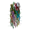
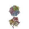
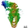
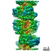
Journal: IUCrJ / Year: 2020
Title: Protein-lipid architecture of a cholinergic postsynaptic membrane.
Authors: Nigel Unwin /
Abstract: The cholinergic postsynaptic membrane is an acetyl-choline receptor-rich membrane mediating fast chemical communication at the nerve-muscle synapse. Here, cryo-EM is used to examine the protein-lipid ...The cholinergic postsynaptic membrane is an acetyl-choline receptor-rich membrane mediating fast chemical communication at the nerve-muscle synapse. Here, cryo-EM is used to examine the protein-lipid architecture of this membrane in tubular vesicles obtained from the (muscle-derived) electric organ of the ray. As reported earlier, the helical arrangement of the protein component of the vesicles facilitates image averaging and enables us to determine how cholesterol and phospho-lipid molecules are distributed in the surrounding matrix, using headgroup size as a means to discriminate between the two kinds of lipid. It is shown that cholesterol segregates preferentially around the receptors in both leaflets of the lipid bilayer, interacting robustly with specific transmembrane sites and creating a network of bridging microdomains. Cholesterol interactions with the receptor are apparently essential for stabilizing and maintaining its physiological architecture, since the transmembrane structure contracts, involving displacements of the helices at the outer membrane surface by ∼2 Å (1-3 Å), when this lipid is extracted. The microdomains may promote cooperativity between neighbouring receptors, leading to an enhanced postsynaptic response.
History
DepositionJun 29, 2020-
Header (metadata) releaseSep 16, 2020-
Map releaseSep 16, 2020-
UpdateSep 16, 2020-
Current statusSep 16, 2020Processing site: PDBe / Status: Released

-
Structure visualization

Movie
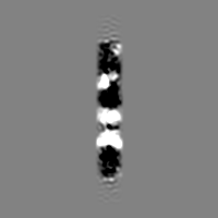
  • Surface view with section colored by density value
  • Surface level: 0.014
  • Imaged by UCSF Chimera
  • Download
  • Surface view colored by cylindrical radius
  • Surface level: 0.014
  • Imaged by UCSF Chimera
  • Download
Movie viewer
Structure viewerEM map:
SurfViewMolmilJmol/JSmol
Supplemental images

Downloads & links

-
Map

FileDownload / File: emd_11239.map.gz / Format: CCP4 / Size: 30.5 MB / Type: IMAGE STORED AS FLOATING POINT NUMBER (4 BYTES)
AnnotationAverage map of membrane-bound resting-state Torpedo nicotinic acetylcholine receptor, obtained from 2 helical families ((-16,6) and (-17,5))
Voxel sizeX=Y=Z: 1.34 Å
Density
Contour LevelBy AUTHOR: 0.014 / Movie #1: 0.014
Minimum - Maximum-0.017447364 - 0.046507176
Average (Standard dev.)0.00027804924 (±0.0024836066)
SymmetrySpace group: 1
Details

EMDB XML:

Map geometry
Axis orderXYZ
Origin000
Dimensions200200200
Spacing200200200
CellA=B=C: 268.0 Å
α=β=γ: 90.0 °

CCP4 map header:

modeImage stored as Reals
Å/pix. X/Y/Z1.341.341.34
M x/y/z200200200
origin x/y/z0.0000.0000.000
length x/y/z268.000268.000268.000
α/β/γ90.00090.00090.000
MAP C/R/S123
start NC/NR/NS000
NC/NR/NS200200200
D min/max/mean-0.0170.0470.000

-
Supplemental data

-
Sample components

-
Entire : Nicotinic acetylcholine receptor

EntireName: Nicotinic acetylcholine receptor
Components
  • Complex: Nicotinic acetylcholine receptor

-
Supramolecule #1: Nicotinic acetylcholine receptor

SupramoleculeName: Nicotinic acetylcholine receptor / type: complex / ID: 1 / Parent: 0
Details: Heteropentamric ion channel comprised of two alpha subunits, beta, gamma and delta
Source (natural)Organism: Torpedo marmorata (marbled electric ray) / Organ: electric organ / Tissue: modified muscle tissue / Organelle: postsynaptic membrane / Location in cell: synapse
Molecular weightTheoretical: 290 KDa

-
Experimental details

-
Structure determination

Methodcryo EM
Processinghelical reconstruction
Aggregation statehelical array

-
Sample preparation

BufferpH: 7 / Component - Concentration: 100.0 mM / Component - Formula: (CH3)2AsO2Na / Component - Name: sodium cacodylate / Details: Solution contains 1mM CaCl2
GridModel: Quantifoil / Material: COPPER / Mesh: 400 / Support film - Material: CARBON / Support film - topology: HOLEY / Support film - Film thickness: 20.0 nm / Pretreatment - Type: GLOW DISCHARGE / Pretreatment - Atmosphere: OTHER
VitrificationCryogen name: ETHANE / Chamber humidity: 95 % / Chamber temperature: 282 K / Instrument: HOMEMADE PLUNGER
Detailsmembrane tubes with acetylcholine receptors arranged on a helical surface lattice

-
Electron microscopy

MicroscopeFEI TITAN KRIOS
TemperatureMin: 70.0 K / Max: 70.0 K
Specialist opticsPhase plate: OTHER
Image recordingFilm or detector model: FEI FALCON III (4k x 4k) / Detector mode: INTEGRATING / Digitization - Dimensions - Width: 4096 pixel / Digitization - Dimensions - Height: 4096 pixel / Digitization - Sampling interval: 14.0 µm / Number grids imaged: 100 / Number real images: 2411 / Average exposure time: 2.0 sec. / Average electron dose: 40.0 e/Å2 / Details: 79 frames per micrograph
Electron beamAcceleration voltage: 300 kV / Electron source: FIELD EMISSION GUN
Electron opticsC2 aperture diameter: 70.0 µm / Calibrated defocus max: 2.8 µm / Calibrated defocus min: 1.0 µm / Calibrated magnification: 59000 / Illumination mode: FLOOD BEAM / Imaging mode: BRIGHT FIELD / Cs: 2.7 mm / Nominal defocus max: 2.8 µm / Nominal defocus min: 1.0 µm / Nominal magnification: 59000
Sample stageSpecimen holder model: FEI TITAN KRIOS AUTOGRID HOLDER / Cooling holder cryogen: NITROGEN
Experimental equipment
Model: Titan Krios / Image courtesy: FEI Company

+
Image processing

Details
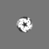
This map is simply an average of two maps of the protein derived from two families of helical membrane tubes. (-16,6) tubes: 1037 images, 51921 segments (-17,5) tubes: 1374 images, 71741 segments
Final reconstructionApplied symmetry - Point group: C1 (asymmetric) / Algorithm: FOURIER SPACE / Resolution.type: BY AUTHOR / Resolution: 5.8 Å / Resolution method: FSC 0.143 CUT-OFF / Software - Name: RELION (ver. 2.1 and 3.0)
Details: FSC is based on comparison of aligned particles cut out from the the two individual helical reconstructions
Number images used: 123662
CTF correctionSoftware - Name: Gctf
Details: CTF amplitude correction was applied locally along the axis of each tube, using the non dose-weighted images
Segment selectionNumber selected: 123662
Final angle assignmentType: NOT APPLICABLE

+
About Yorodumi

-
News

-
Feb 9, 2022. New format data for meta-information of EMDB entries

New format data for meta-information of EMDB entries

  • Version 3 of the EMDB header file is now the official format.
  • The previous official version 1.9 will be removed from the archive.

Related info.:EMDB header

External links:wwPDB to switch to version 3 of the EMDB data model

-
Aug 12, 2020. Covid-19 info

Covid-19 info

URL: https://pdbjlvh1.pdbj.org/emnavi/covid19.php

New page: Covid-19 featured information page in EM Navigator.

Related info.:Covid-19 info / Mar 5, 2020. Novel coronavirus structure data

+
Mar 5, 2020. Novel coronavirus structure data

Novel coronavirus structure data

Related info.:Yorodumi Speices / Aug 12, 2020. Covid-19 info

External links:COVID-19 featured content - PDBj / Molecule of the Month (242):Coronavirus Proteases

+
Jan 31, 2019. EMDB accession codes are about to change! (news from PDBe EMDB page)

EMDB accession codes are about to change! (news from PDBe EMDB page)

  • The allocation of 4 digits for EMDB accession codes will soon come to an end. Whilst these codes will remain in use, new EMDB accession codes will include an additional digit and will expand incrementally as the available range of codes is exhausted. The current 4-digit format prefixed with “EMD-” (i.e. EMD-XXXX) will advance to a 5-digit format (i.e. EMD-XXXXX), and so on. It is currently estimated that the 4-digit codes will be depleted around Spring 2019, at which point the 5-digit format will come into force.
  • The EM Navigator/Yorodumi systems omit the EMD- prefix.

Related info.:Q: What is EMD? / ID/Accession-code notation in Yorodumi/EM Navigator

External links:EMDB Accession Codes are Changing Soon! / Contact to PDBj

+
Jul 12, 2017. Major update of PDB

Major update of PDB

  • wwPDB released updated PDB data conforming to the new PDBx/mmCIF dictionary.
  • This is a major update changing the version number from 4 to 5, and with Remediation, in which all the entries are updated.
  • In this update, many items about electron microscopy experimental information are reorganized (e.g. em_software).
  • Now, EM Navigator and Yorodumi are based on the updated data.

External links:wwPDB Remediation / Enriched Model Files Conforming to OneDep Data Standards Now Available in the PDB FTP Archive

-
Yorodumi

Thousand views of thousand structures

  • Yorodumi is a browser for structure data from EMDB, PDB, SASBDB, etc.
  • This page is also the successor to EM Navigator detail page, and also detail information page/front-end page for Omokage search.
  • The word "yorodu" (or yorozu) is an old Japanese word meaning "ten thousand". "mi" (miru) is to see.

Related info.:EMDB / PDB / SASBDB / Comparison of 3 databanks / Yorodumi Search / Aug 31, 2016. New EM Navigator & Yorodumi / Yorodumi Papers / Jmol/JSmol / Function and homology information / Changes in new EM Navigator and Yorodumi

Read more