Engineering and Physical Sciences Research Council
EP/S025243/1
United Kingdom
Wellcome Trust
100209/Z/12/Z
United Kingdom
Cancer Research UK
C20724/A14414
United Kingdom
Cancer Research UK
C20724/A26752
United Kingdom
Wellcome Trust
101122/Z/13/Z
United Kingdom
Medical Research Council (MRC, United Kingdom)
MR/N00065X/1
United Kingdom
Citation
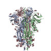
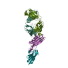
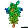
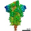
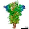
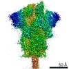
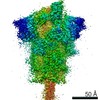
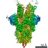
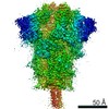
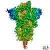
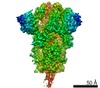
Journal: Nat Struct Mol Biol / Year: 2020 Title: Neutralizing nanobodies bind SARS-CoV-2 spike RBD and block interaction with ACE2. Authors: Jiandong Huo / Audrey Le Bas / Reinis R Ruza / Helen M E Duyvesteyn / Halina Mikolajek / Tomas Malinauskas / Tiong Kit Tan / Pramila Rijal / Maud Dumoux / Philip N Ward / Jingshan Ren / ...Authors: Jiandong Huo / Audrey Le Bas / Reinis R Ruza / Helen M E Duyvesteyn / Halina Mikolajek / Tomas Malinauskas / Tiong Kit Tan / Pramila Rijal / Maud Dumoux / Philip N Ward / Jingshan Ren / Daming Zhou / Peter J Harrison / Miriam Weckener / Daniel K Clare / Vinod K Vogirala / Julika Radecke / Lucile Moynié / Yuguang Zhao / Javier Gilbert-Jaramillo / Michael L Knight / Julia A Tree / Karen R Buttigieg / Naomi Coombes / Michael J Elmore / Miles W Carroll / Loic Carrique / Pranav N M Shah / William James / Alain R Townsend / David I Stuart / Raymond J Owens / James H Naismith / Abstract: The SARS-CoV-2 virus is more transmissible than previous coronaviruses and causes a more serious illness than influenza. The SARS-CoV-2 receptor binding domain (RBD) of the spike protein binds to the ...The SARS-CoV-2 virus is more transmissible than previous coronaviruses and causes a more serious illness than influenza. The SARS-CoV-2 receptor binding domain (RBD) of the spike protein binds to the human angiotensin-converting enzyme 2 (ACE2) receptor as a prelude to viral entry into the cell. Using a naive llama single-domain antibody library and PCR-based maturation, we have produced two closely related nanobodies, H11-D4 and H11-H4, that bind RBD (K of 39 and 12 nM, respectively) and block its interaction with ACE2. Single-particle cryo-EM revealed that both nanobodies bind to all three RBDs in the spike trimer. Crystal structures of each nanobody-RBD complex revealed how both nanobodies recognize the same epitope, which partly overlaps with the ACE2 binding surface, explaining the blocking of the RBD-ACE2 interaction. Nanobody-Fc fusions showed neutralizing activity against SARS-CoV-2 (4-6 nM for H11-H4, 18 nM for H11-D4) and additive neutralization with the SARS-CoV-1/2 antibody CR3022.
History
Deposition
May 22, 2020
-
Header (metadata) release
Jun 3, 2020
-
Map release
Jun 3, 2020
-
Update
Oct 16, 2024
-
Current status
Oct 16, 2024
Processing site: PDBe / Status: Released
-
Structure visualization
Movie
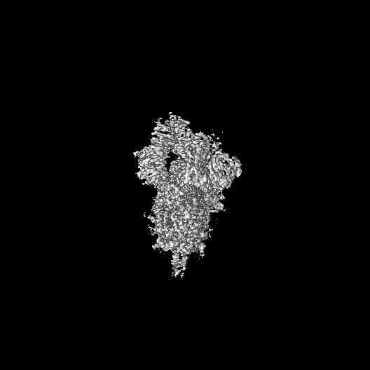
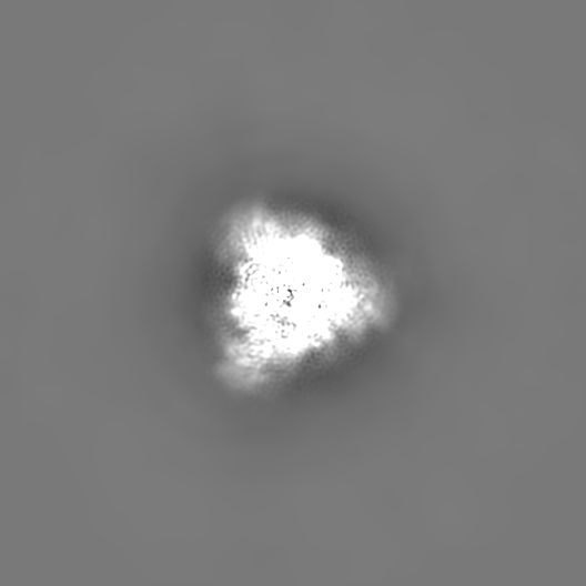
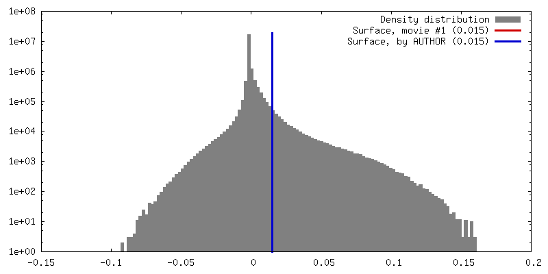
Surface view with section colored by density value
Model: C-flat-2/1 / Material: COPPER / Mesh: 200 / Support film - Material: CARBON / Support film - topology: HOLEY / Pretreatment - Type: GLOW DISCHARGE / Pretreatment - Time: 20 sec. / Pretreatment - Atmosphere: AIR
Vitrification
Cryogen name: ETHANE / Chamber humidity: 100 % / Chamber temperature: 277.65 K / Instrument: FEI VITROBOT MARK IV / Details: Blot for 6 seconds with a blotting force of -1.
-
Electron microscopy
Microscope
FEI TITAN KRIOS
Image recording
Film or detector model: GATAN K3 (6k x 4k) / Average electron dose: 42.8 e/Å2
Electron beam
Acceleration voltage: 300 kV / Electron source: FIELD EMISSION GUN
Electron optics
Illumination mode: FLOOD BEAM / Imaging mode: BRIGHT FIELD
In the structure databanks used in Yorodumi, some data are registered as the other names, "COVID-19 virus" and "2019-nCoV". Here are the details of the virus and the list of structure data.
Jan 31, 2019. EMDB accession codes are about to change! (news from PDBe EMDB page)
EMDB accession codes are about to change! (news from PDBe EMDB page)
The allocation of 4 digits for EMDB accession codes will soon come to an end. Whilst these codes will remain in use, new EMDB accession codes will include an additional digit and will expand incrementally as the available range of codes is exhausted. The current 4-digit format prefixed with “EMD-” (i.e. EMD-XXXX) will advance to a 5-digit format (i.e. EMD-XXXXX), and so on. It is currently estimated that the 4-digit codes will be depleted around Spring 2019, at which point the 5-digit format will come into force.
The EM Navigator/Yorodumi systems omit the EMD- prefix.
Related info.:Q: What is EMD? / ID/Accession-code notation in Yorodumi/EM Navigator
Yorodumi is a browser for structure data from EMDB, PDB, SASBDB, etc.
This page is also the successor to EM Navigator detail page, and also detail information page/front-end page for Omokage search.
The word "yorodu" (or yorozu) is an old Japanese word meaning "ten thousand". "mi" (miru) is to see.
Related info.:EMDB / PDB / SASBDB / Comparison of 3 databanks / Yorodumi Search / Aug 31, 2016. New EM Navigator & Yorodumi / Yorodumi Papers / Jmol/JSmol / Function and homology information / Changes in new EM Navigator and Yorodumi
 Movie
Movie Controller

 Yorodumi
 Open data
 Basic information
 Map data
 Sample
 Keywords
 Function and homology information
 Severe acute respiratory syndrome coronavirus 2 /
 Authors
 United Kingdom, 6 items
  Citation
 Structure visualization
 Downloads & links
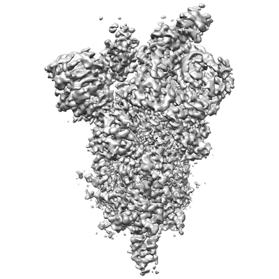 emd_11068.png
 http://ftp.pdbj.org/pub/emdb/structures/EMD-11068
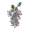


 Z (Sec.)
 Y (Row.)
 X (Col.)


 Sample components
 Homo sapiens (human)

 Processing
 Electron microscopy
 FIELD EMISSION GUN