Movie
Movie Controller

[English] 日本語
 Yorodumi
- EMDB-11046: In situ cryo-electron tomography reveals layered organization of ... -
+ Open data

- Basic information
| Entry | Database: EMDB / ID: EMD-11046 | |||||||||
|---|---|---|---|---|---|---|---|---|---|---|
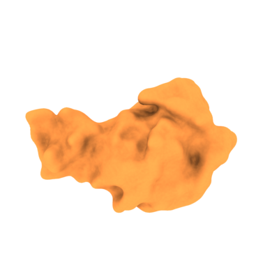
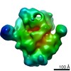
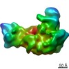
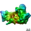
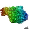
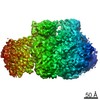
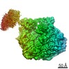
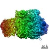
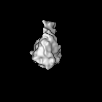
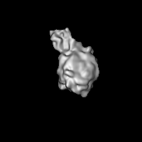
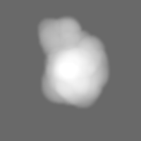
| Title | In situ cryo-electron tomography reveals layered organization of pre-ribosome maturation in nucleoli. Large Subunit Pre-Ribosome, Class 1 | |||||||||
 Map data Map data | Chlamydomonas reinhardtii large subunit pre-ribosome, Class 1 | |||||||||
 Sample Sample |
| |||||||||
| Biological species |   Chlamydomonas reinhardtii (plant) Chlamydomonas reinhardtii (plant) | |||||||||
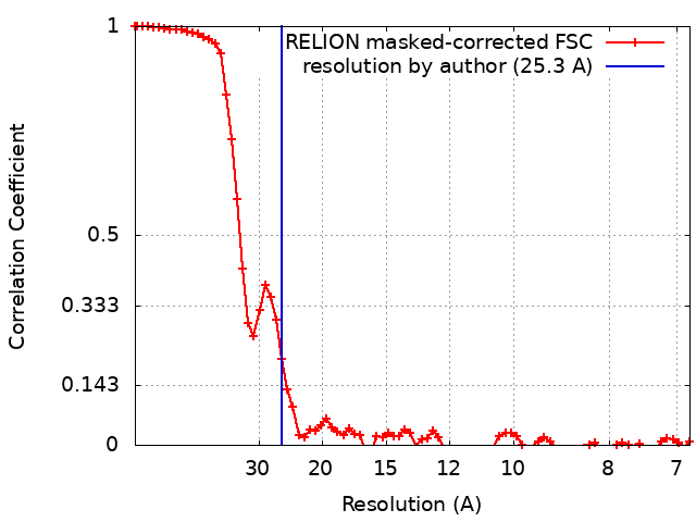
| Method | subtomogram averaging / cryo EM / Resolution: 25.3 Å | |||||||||
 Authors Authors | Erdmann PS / Klumpe S / Hou Z / Beck F / Wilfling F / Plitzko JM / Baumeister W | |||||||||
| Funding support |  Germany, 1 items Germany, 1 items
| |||||||||
 Citation Citation | Journal: Nat Commun / Year: 2021 Title: In situ cryo-electron tomography reveals gradient organization of ribosome biogenesis in intact nucleoli. Authors: Philipp S Erdmann / Zhen Hou / Sven Klumpe / Sagar Khavnekar / Florian Beck / Florian Wilfling / Jürgen M Plitzko / Wolfgang Baumeister /  Abstract: Ribosomes comprise a large (LSU) and a small subunit (SSU) which are synthesized independently in the nucleolus before being exported into the cytoplasm, where they assemble into functional ribosomes. ...Ribosomes comprise a large (LSU) and a small subunit (SSU) which are synthesized independently in the nucleolus before being exported into the cytoplasm, where they assemble into functional ribosomes. Individual maturation steps have been analyzed in detail using biochemical methods, light microscopy and conventional electron microscopy (EM). In recent years, single particle analysis (SPA) has yielded molecular resolution structures of several pre-ribosomal intermediates. It falls short, however, of revealing the spatiotemporal sequence of ribosome biogenesis in the cellular context. Here, we present our study on native nucleoli in Chlamydomonas reinhardtii, in which we follow the formation of LSU and SSU precursors by in situ cryo-electron tomography (cryo-ET) and subtomogram averaging (STA). By combining both positional and molecular data, we reveal gradients of ribosome maturation within the granular component (GC), offering a new perspective on how the liquid-liquid-phase separation of the nucleolus supports ribosome biogenesis. | |||||||||
| History |
|
- Structure visualization
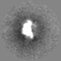
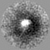
| Movie |
 Movie viewer Movie viewer |
|---|---|
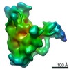
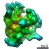
| Structure viewer | EM map: SurfViewMolmilJmol/JSmol |
| Supplemental images |
- Downloads & links
-EMDB archive
| Map data | emd_11046.map.gz | 28.2 MB | EMDB map data format | |
|---|---|---|---|---|
| Header (meta data) | emd-11046-v30.xmlemd-11046.xml | 11.4 KB 11.4 KB | Display Display | EMDB header |
| FSC (resolution estimation) | emd_11046_fsc.xml | 7.3 KB | Display | FSC data file |
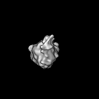
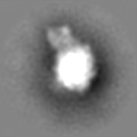
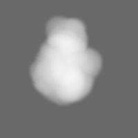
| Images |  emd_11046.png emd_11046.png | 46.7 KB | ||
| Masks | emd_11046_msk_1.map | 30.5 MB | Mask map | |
| Archive directory |  http://ftp.pdbj.org/pub/emdb/structures/EMD-11046ftp://ftp.pdbj.org/pub/emdb/structures/EMD-11046 http://ftp.pdbj.org/pub/emdb/structures/EMD-11046ftp://ftp.pdbj.org/pub/emdb/structures/EMD-11046 | HTTPS FTP |
-Validation report
| Summary document | emd_11046_validation.pdf.gz | 351.8 KB | Display | EMDB validaton report |
|---|---|---|---|---|
| Full document | emd_11046_full_validation.pdf.gz | 351.4 KB | Display | |
| Data in XML | emd_11046_validation.xml.gz | 9.8 KB | Display | |
| Data in CIF | emd_11046_validation.cif.gz | 12.6 KB | Display | |
| Arichive directory | https://ftp.pdbj.org/pub/emdb/validation_reports/EMD-11046ftp://ftp.pdbj.org/pub/emdb/validation_reports/EMD-11046 | HTTPS FTP |
-Related structure data
| Related structure data | C: citing same article ( |
|---|---|
| Similar structure data |


-Links
| EMDB pages | EMDB (EBI/PDBe) / EMDataResource |
|---|---|
| Related items in Molecule of the Month |

-Map
| File | Download / File: emd_11046.map.gz / Format: CCP4 / Size: 30.5 MB / Type: IMAGE STORED AS FLOATING POINT NUMBER (4 BYTES) | ||||||||||||||||||||||||||||||||||||||||||||||||||||||||||||
|---|---|---|---|---|---|---|---|---|---|---|---|---|---|---|---|---|---|---|---|---|---|---|---|---|---|---|---|---|---|---|---|---|---|---|---|---|---|---|---|---|---|---|---|---|---|---|---|---|---|---|---|---|---|---|---|---|---|---|---|---|---|
| Annotation | Chlamydomonas reinhardtii large subunit pre-ribosome, Class 1 | ||||||||||||||||||||||||||||||||||||||||||||||||||||||||||||
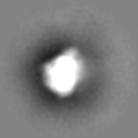
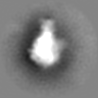
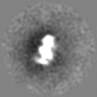
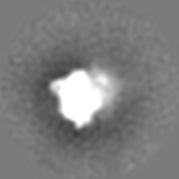
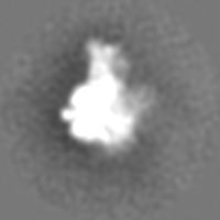
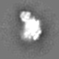
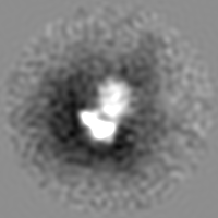
| Projections & slices | Image control
Images are generated by Spider. | ||||||||||||||||||||||||||||||||||||||||||||||||||||||||||||
| Voxel size | X=Y=Z: 3.42 Å | ||||||||||||||||||||||||||||||||||||||||||||||||||||||||||||
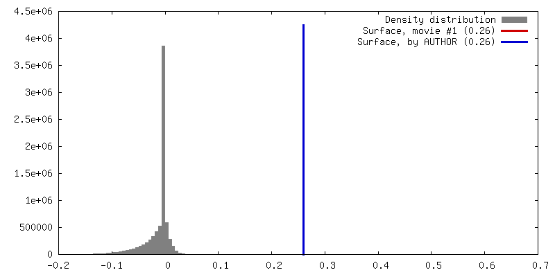
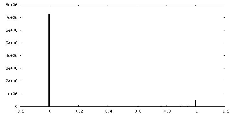
| Density |
| ||||||||||||||||||||||||||||||||||||||||||||||||||||||||||||
| Symmetry | Space group: 1 | ||||||||||||||||||||||||||||||||||||||||||||||||||||||||||||
| Details | EMDB XML:
CCP4 map header:
| ||||||||||||||||||||||||||||||||||||||||||||||||||||||||||||
 Z (Sec.)
 Y (Row.)
 X (Col.)


-Supplemental data
-Mask #1
| File | emd_11046_msk_1.map | ||||||||||||
|---|---|---|---|---|---|---|---|---|---|---|---|---|---|
| Projections & Slices |
| ||||||||||||
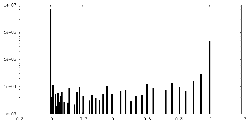
| Density Histograms |


- Sample components
-Entire : nucleolar small subunit processome, class 1
| Entire | Name: nucleolar small subunit processome, class 1 |
|---|---|
| Components |
|
-Supramolecule #1: nucleolar small subunit processome, class 1
| Supramolecule | Name: nucleolar small subunit processome, class 1 / type: complex / ID: 1 / Parent: 0 |
|---|---|
| Source (natural) | Organism: Chlamydomonas reinhardtii (plant) / Location in cell: nuclear |
-Experimental details
-Structure determination
| Method | cryo EM |
|---|---|
 Processing Processing | subtomogram averaging |
| Aggregation state | particle |
-Sample preparation
| Buffer | pH: 7.4 |
|---|---|
| Vitrification | Cryogen name: ETHANE-PROPANE / Chamber humidity: 100 % / Chamber temperature: 298 K / Instrument: FEI VITROBOT MARK IV |
| Details | in situ focus ion beam milled cells |
- Electron microscopy
| Microscope | FEI TITAN KRIOS |
|---|---|
| Specialist optics | Energy filter - Name: GIF Bioquantum / Energy filter - Slit width: 20 eV |
| Image recording | Film or detector model: GATAN K2 SUMMIT (4k x 4k) / Detector mode: COUNTING / Average electron dose: 1.5 e/Å2 Details: mixture of dose-symmetric, and bidirectional tilting schemes |
| Electron beam | Acceleration voltage: 300 kV / Electron source:  FIELD EMISSION GUN FIELD EMISSION GUN |
| Electron optics | Illumination mode: FLOOD BEAM / Imaging mode: BRIGHT FIELD |
| Experimental equipment |  Model: Titan Krios / Image courtesy: FEI Company |