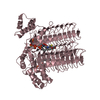
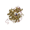
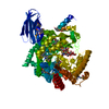
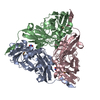
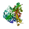
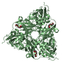
Journal: mBio / Year: 2020 Title: Isolated Heme A Synthase from Aquifex aeolicus Is a Trimer. Authors: Hui Zeng / Guoliang Zhu / Shuangbo Zhang / Xinmei Li / Janosch Martin / Nina Morgner / Fei Sun / Guohong Peng / Hao Xie / Hartmut Michel / Abstract: The integral membrane protein heme A synthase (HAS) catalyzes the biosynthesis of heme A, which is a prerequisite for cellular respiration in a wide range of aerobic organisms. Previous studies have ...The integral membrane protein heme A synthase (HAS) catalyzes the biosynthesis of heme A, which is a prerequisite for cellular respiration in a wide range of aerobic organisms. Previous studies have revealed that HAS can form homo-oligomeric complexes, and this oligomerization appears to be evolutionarily conserved among prokaryotes and eukaryotes and is shown to be essential for the biological function of eukaryotic HAS. Despite its importance, little is known about the detailed structural properties of HAS oligomers. Here, we aimed to address this critical issue by analyzing the oligomeric state of HAS from (AaHAS) using a combination of techniques, including size exclusion chromatography coupled with multiangle light scattering (SEC-MALS), cross-linking, laser-induced liquid bead ion desorption mass spectrometry (LILBID-MS), and single-particle electron cryomicroscopy (cryo-EM). Our results show that HAS forms a thermostable trimeric complex. A cryo-EM density map provides information on the oligomerization interface of the AaHAS trimer. These results provide structural insights into HAS multimerization and expand our knowledge of this important enzyme. Heme A is a vital redox cofactor unique for the terminal cytochrome oxidase in mitochondria and many microorganisms. It plays a key role in oxygen reduction by serving as an electron carrier and as the oxygen-binding site. Heme A is synthesized from heme O by an integral membrane protein, heme A synthase (HAS). Defects in HAS impair cellular respiration and have been linked to various human diseases, e.g., fatal infantile hypertrophic cardiomyopathy and Leigh syndrome. HAS exists as a stable oligomeric complex, and studies have shown that oligomerization of eukaryotic HAS is necessary for its proper function. However, the molecular architecture of the HAS oligomeric complex has remained uncharacterized. The present study shows that HAS forms trimers and reveals how the oligomeric arrangement contributes to the complex stability and flexibility, enabling HAS to perform its catalytic function effectively. This work provides the basic understanding for future studies on heme A biosynthesis.
History
Deposition
Apr 30, 2020
-
Header (metadata) release
Jun 17, 2020
-
Map release
Jun 17, 2020
-
Update
Jun 17, 2020
-
Current status
Jun 17, 2020
Processing site: PDBe / Status: Released
-
Structure visualization
Movie
Surface view with section colored by density value
In the structure databanks used in Yorodumi, some data are registered as the other names, "COVID-19 virus" and "2019-nCoV". Here are the details of the virus and the list of structure data.
Jan 31, 2019. EMDB accession codes are about to change! (news from PDBe EMDB page)
EMDB accession codes are about to change! (news from PDBe EMDB page)
The allocation of 4 digits for EMDB accession codes will soon come to an end. Whilst these codes will remain in use, new EMDB accession codes will include an additional digit and will expand incrementally as the available range of codes is exhausted. The current 4-digit format prefixed with “EMD-” (i.e. EMD-XXXX) will advance to a 5-digit format (i.e. EMD-XXXXX), and so on. It is currently estimated that the 4-digit codes will be depleted around Spring 2019, at which point the 5-digit format will come into force.
The EM Navigator/Yorodumi systems omit the EMD- prefix.
Related info.:Q: What is EMD? / ID/Accession-code notation in Yorodumi/EM Navigator
Yorodumi is a browser for structure data from EMDB, PDB, SASBDB, etc.
This page is also the successor to EM Navigator detail page, and also detail information page/front-end page for Omokage search.
The word "yorodu" (or yorozu) is an old Japanese word meaning "ten thousand". "mi" (miru) is to see.
Related info.:EMDB / PDB / SASBDB / Comparison of 3 databanks / Yorodumi Search / Aug 31, 2016. New EM Navigator & Yorodumi / Yorodumi Papers / Jmol/JSmol / Function and homology information / Changes in new EM Navigator and Yorodumi
 Movie
Movie Controller


 Open data
 Basic information
 Map data
 Sample
 Aquifex aeolicus VF5 (bacteria)
 Authors
 Citation

 Structure visualization
 Movie viewer
 Downloads & links
 emd_10987.png
 http://ftp.pdbj.org/pub/emdb/structures/EMD-10987
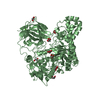

 Z (Sec.)
 Y (Row.)
 X (Col.)


 Sample components
 Processing
 Electron microscopy
 FIELD EMISSION GUN

