+ Open data
- Basic information
| Entry | Database: EMDB / ID: EMD-1098 | |||||||||
|---|---|---|---|---|---|---|---|---|---|---|
| Title | Three-dimensional structure of a bacterial oxalate transporter. | |||||||||
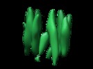
 Map data Map data | This is an image of the bacterial oxalate transporter(OxlT)in the oxalate-bound, "closed" state. OxlT is a representative member of the Major Facilitator Superfamily (MFS). | |||||||||
 Sample Sample |
| |||||||||
| Biological species |  Oxalobacter formigenes (bacteria) Oxalobacter formigenes (bacteria) | |||||||||
| Method | electron crystallography / cryo EM / Resolution: 6.5 Å | |||||||||
 Authors Authors | Hirai T / Heymann JAW / Shi D / Sarker R / Maloney PC / Subramaniam S | |||||||||
 Citation Citation |  Journal: Nat Struct Biol / Year: 2002 Journal: Nat Struct Biol / Year: 2002Title: Three-dimensional structure of a bacterial oxalate transporter. Authors: Teruhisa Hirai / Jürgen A W Heymann / Dan Shi / Rafiquel Sarker / Peter C Maloney / Sriram Subramaniam /  Abstract: The major facilitator superfamily (MFS) represents one of the largest classes of evolutionarily related membrane transporter proteins. Here we present the three-dimensional structure at 6.5 A ...The major facilitator superfamily (MFS) represents one of the largest classes of evolutionarily related membrane transporter proteins. Here we present the three-dimensional structure at 6.5 A resolution of a bacterial member of this superfamily, OxlT. The structure, derived from an electron crystallographic analysis of two-dimensional crystals, reveals that the 12 helices in the OxlT molecule are arranged around a central cavity, which is widest at the center of the membrane. The helices divide naturally into three groups: a peripheral set comprising helices 3, 6, 9 and 12; a second set comprising helices 2, 5, 8 and 11 that faces the central substrate transport pathway across most of the length of the membrane; and a third set comprising helices 1, 4, 7 and 10 that participate in the pathway either on the cytoplasmic side (4 and 10) or on the periplasmic side (1 and 7). Overall, the architecture of the protein is remarkably symmetric, providing a compelling molecular explanation for the ability of such transporters to carry out bi-directional substrate transport. | |||||||||
| History |
|
- Structure visualization
| Movie |
 Movie viewer Movie viewer |
|---|---|
| Structure viewer | EM map:  SurfView SurfView Molmil Molmil Jmol/JSmol Jmol/JSmol |
| Supplemental images |
- Downloads & links
-EMDB archive
| Map data |  emd_1098.map.gz emd_1098.map.gz | 3.1 MB |  EMDB map data format EMDB map data format | |
|---|---|---|---|---|
| Header (meta data) |  emd-1098-v30.xml emd-1098-v30.xml emd-1098.xml emd-1098.xml | 10.9 KB 10.9 KB | Display Display |  EMDB header EMDB header |
| Images |  1098.gif 1098.gif | 14.8 KB | ||
| Archive directory |  http://ftp.pdbj.org/pub/emdb/structures/EMD-1098 http://ftp.pdbj.org/pub/emdb/structures/EMD-1098 ftp://ftp.pdbj.org/pub/emdb/structures/EMD-1098 ftp://ftp.pdbj.org/pub/emdb/structures/EMD-1098 | HTTPS FTP |
- Links
Links
| EMDB pages |  EMDB (EBI/PDBe) / EMDB (EBI/PDBe) /  EMDataResource EMDataResource |
|---|
- Map
Map
| File |  Download / File: emd_1098.map.gz / Format: CCP4 / Size: 3.3 MB / Type: IMAGE STORED AS FLOATING POINT NUMBER (4 BYTES) Download / File: emd_1098.map.gz / Format: CCP4 / Size: 3.3 MB / Type: IMAGE STORED AS FLOATING POINT NUMBER (4 BYTES) | ||||||||||||||||||||||||||||||||||||||||||||||||||||||||||||||||||||
|---|---|---|---|---|---|---|---|---|---|---|---|---|---|---|---|---|---|---|---|---|---|---|---|---|---|---|---|---|---|---|---|---|---|---|---|---|---|---|---|---|---|---|---|---|---|---|---|---|---|---|---|---|---|---|---|---|---|---|---|---|---|---|---|---|---|---|---|---|---|
| Annotation | This is an image of the bacterial oxalate transporter(OxlT)in the oxalate-bound, "closed" state. OxlT is a representative member of the Major Facilitator Superfamily (MFS). | ||||||||||||||||||||||||||||||||||||||||||||||||||||||||||||||||||||
| Projections & slices | Image control
Images are generated by Spider. generated in cubic-lattice coordinate | ||||||||||||||||||||||||||||||||||||||||||||||||||||||||||||||||||||
| Voxel size | X: 0.5511 Å / Y: 0.52667 Å / Z: 0.54945 Å | ||||||||||||||||||||||||||||||||||||||||||||||||||||||||||||||||||||
| Density |
| ||||||||||||||||||||||||||||||||||||||||||||||||||||||||||||||||||||
| Symmetry | Space group: 18 | ||||||||||||||||||||||||||||||||||||||||||||||||||||||||||||||||||||
| Details | EMDB XML:
CCP4 map header:
| ||||||||||||||||||||||||||||||||||||||||||||||||||||||||||||||||||||
-Supplemental data
- Sample components
-Entire : Oxalate Transporter
| Entire | Name: Oxalate Transporter |
|---|---|
| Components |
|
-Supramolecule #1000: Oxalate Transporter
| Supramolecule | Name: Oxalate Transporter / type: sample / ID: 1000 / Number unique components: 1 |
|---|
-Macromolecule #1: oxalate transporter
| Macromolecule | Name: oxalate transporter / type: protein_or_peptide / ID: 1 / Name.synonym: OxlT Details: Oxalate(-ooc-coo-) is believed to be bound in the central cavity of OxlT in this crystal form. Endogenous E.coli lipids and added synthetic lipids are expected to be present in this two ...Details: Oxalate(-ooc-coo-) is believed to be bound in the central cavity of OxlT in this crystal form. Endogenous E.coli lipids and added synthetic lipids are expected to be present in this two dimensional crystal. No detectable density for lipid or oxalate is observed at current resolution. Recombinant expression: Yes |
|---|---|
| Source (natural) | Organism:  Oxalobacter formigenes (bacteria) / Location in cell: cell membrane Oxalobacter formigenes (bacteria) / Location in cell: cell membrane |
| Molecular weight | Experimental: 44 KDa |
| Recombinant expression | Organism:  |
-Experimental details
-Structure determination
| Method | cryo EM |
|---|---|
 Processing Processing | electron crystallography |
| Aggregation state | 2D array |
- Sample preparation
| Grid | Details: 400 mesh Cu grid |
|---|---|
| Vitrification | Cryogen name: NITROGEN / Chamber temperature: 77 K / Instrument: HOMEMADE PLUNGER / Details: Vitrification instrument: MRC plunger |
- Electron microscopy
| Microscope | FEI TECNAI F30 |
|---|---|
| Temperature | Min: 90 K / Max: 95 K |
| Alignment procedure | Legacy - Astigmatism: astigmatism was corrected at 200,000 times magnification |
| Details | Tecnai12, 120KV with tungsten filament was also used at early stage. Floodbeam was also used for some images as illumination mode. |
| Image recording | Category: FILM / Film or detector model: KODAK SO-163 FILM / Digitization - Scanner: ZEISS SCAI / Digitization - Sampling interval: 7 µm / Number real images: 47 / Average electron dose: 10 e/Å2 Details: The best images were selected by optical diffraction. Od range: 0.5 / Bits/pixel: 10 |
| Tilt angle min | 0 |
| Electron beam | Acceleration voltage: 300 kV / Electron source:  FIELD EMISSION GUN FIELD EMISSION GUN |
| Electron optics | Calibrated magnification: 57000 / Illumination mode: SPOT SCAN / Imaging mode: BRIGHT FIELD / Cs: 2.2 mm / Nominal defocus max: 2.0 µm / Nominal defocus min: 0.1 µm / Nominal magnification: 59000 |
| Sample stage | Specimen holder: side entry / Specimen holder model: GATAN LIQUID NITROGEN / Tilt angle max: 45 / Tilt series - Axis1 - Min angle: 0 ° / Tilt series - Axis1 - Max angle: 45 ° |
| Experimental equipment |  Model: Tecnai F30 / Image courtesy: FEI Company |
- Image processing
| Details | Specimens were either embedded in 3.5% trehalose or prepared as frozen-hydrated specimens. |
|---|---|
| Final reconstruction | Resolution.type: BY AUTHOR / Resolution: 6.5 Å / Resolution method: OTHER / Software - Name: MRC Details: Amplitudes were scaled with respect to reference amplitudes from the helix model. |
| Crystal parameters | Unit cell - A: 100.3 Å / Unit cell - B: 79 Å / Unit cell - C: 100 Å / Unit cell - γ: 90 ° / Unit cell - α: 90 ° / Unit cell - β: 90 ° / Plane group: P 2 21 21 |
| CTF correction | Details: ctfsearch or ttrefine on each image |
 Movie
Movie Controller


 Z (Sec.)
 Y (Row.)
 X (Col.)


