 Movie
Movie Controller

[English] 日本語
 Yorodumi
- EMDB-10766: Tomogram of a mouse neuron including segmentation of the Golgi ap... -
+ Open data

- Basic information
| Entry | Database: EMDB / ID: EMD-10766 | ||||||||||||
|---|---|---|---|---|---|---|---|---|---|---|---|---|---|
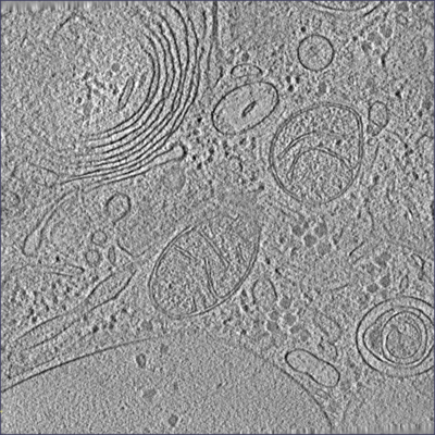
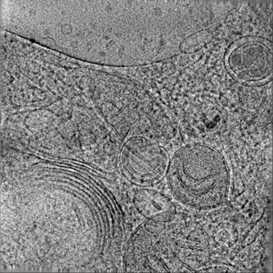
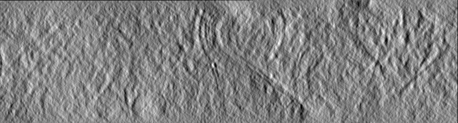
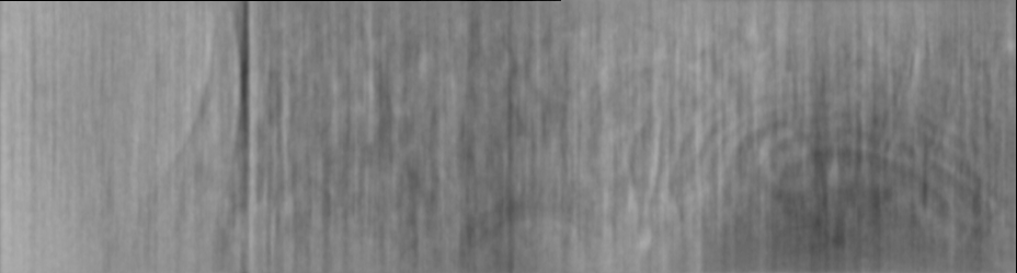
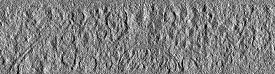
| Title | Tomogram of a mouse neuron including segmentation of the Golgi apparatus and the spherical vesicles for validating a curvature estimation algorithm. | ||||||||||||
 Map data Map data | Raw tomogram. | ||||||||||||
 Sample Sample |
| ||||||||||||
| Biological species |  | ||||||||||||
| Method | electron tomography / cryo EM | ||||||||||||
 Authors Authors | Schaefer T / Salfer M | ||||||||||||
| Funding support |  Germany, 3 items Germany, 3 items
| ||||||||||||
 Citation Citation | Journal: PLoS Comput Biol / Year: 2020 Title: Reliable estimation of membrane curvature for cryo-electron tomography. Authors: Maria Salfer / Javier F Collado / Wolfgang Baumeister / Rubén Fernández-Busnadiego / Antonio Martínez-Sánchez / Abstract: Curvature is a fundamental morphological descriptor of cellular membranes. Cryo-electron tomography (cryo-ET) is particularly well-suited to visualize and analyze membrane morphology in a close-to- ...Curvature is a fundamental morphological descriptor of cellular membranes. Cryo-electron tomography (cryo-ET) is particularly well-suited to visualize and analyze membrane morphology in a close-to-native state and molecular resolution. However, current curvature estimation methods cannot be applied directly to membrane segmentations in cryo-ET, as these methods cannot cope with some of the artifacts introduced during image acquisition and membrane segmentation, such as quantization noise and open borders. Here, we developed and implemented a Python package for membrane curvature estimation from tomogram segmentations, which we named PyCurv. From a membrane segmentation, a signed surface (triangle mesh) is first extracted. The triangle mesh is then represented by a graph, which facilitates finding neighboring triangles and the calculation of geodesic distances necessary for local curvature estimation. PyCurv estimates curvature based on tensor voting. Beside curvatures, this algorithm also provides robust estimations of surface normals and principal directions. We tested PyCurv and three well-established methods on benchmark surfaces and biological data. This revealed the superior performance of PyCurv not only for cryo-ET, but also for data generated by other techniques such as light microscopy and magnetic resonance imaging. Altogether, PyCurv is a versatile open-source software to reliably estimate curvature of membranes and other surfaces in a wide variety of applications. | ||||||||||||
| History |
|
- Structure visualization
| Movie |
 Movie viewer Movie viewer |
|---|---|
| Supplemental images |
- Downloads & links
-EMDB archive
| Map data | emd_10766.map.gz | 310.1 MB | EMDB map data format | |
|---|---|---|---|---|
| Header (meta data) | emd-10766-v30.xmlemd-10766.xml | 12.3 KB 12.3 KB | Display Display | EMDB header |
| Images |  emd_10766.png emd_10766.png | 252.7 KB | ||
| Masks | emd_10766_msk_1.mapemd_10766_msk_2.map | 205.3 MB 205.3 MB | Mask map | |
| Others | emd_10766_additional.map.gzemd_10766_additional_1.map.gz | 392.7 MB 392.7 MB | ||
| Archive directory |  http://ftp.pdbj.org/pub/emdb/structures/EMD-10766ftp://ftp.pdbj.org/pub/emdb/structures/EMD-10766 http://ftp.pdbj.org/pub/emdb/structures/EMD-10766ftp://ftp.pdbj.org/pub/emdb/structures/EMD-10766 | HTTPS FTP |
-Related structure data

-Links
| EMDB pages | EMDB (EBI/PDBe) / EMDataResource |
|---|
-Map
| File | Download / File: emd_10766.map.gz / Format: CCP4 / Size: 410.6 MB / Type: IMAGE STORED AS SIGNED INTEGER (2 BYTES) | ||||||||||||||||||||||||||||||||||||||||||||||||||||||||||||
|---|---|---|---|---|---|---|---|---|---|---|---|---|---|---|---|---|---|---|---|---|---|---|---|---|---|---|---|---|---|---|---|---|---|---|---|---|---|---|---|---|---|---|---|---|---|---|---|---|---|---|---|---|---|---|---|---|---|---|---|---|---|
| Annotation | Raw tomogram. | ||||||||||||||||||||||||||||||||||||||||||||||||||||||||||||
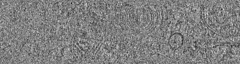
| Projections & slices | Image control
Images are generated by Spider. generated in cubic-lattice coordinate | ||||||||||||||||||||||||||||||||||||||||||||||||||||||||||||
| Voxel size | X=Y=Z: 16.84 Å | ||||||||||||||||||||||||||||||||||||||||||||||||||||||||||||
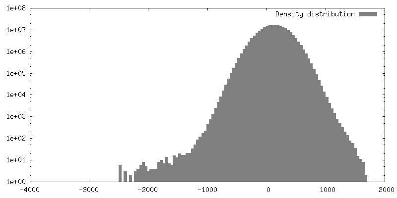
| Density |
| ||||||||||||||||||||||||||||||||||||||||||||||||||||||||||||
| Symmetry | Space group: 1 | ||||||||||||||||||||||||||||||||||||||||||||||||||||||||||||
| Details | EMDB XML:
CCP4 map header:
| ||||||||||||||||||||||||||||||||||||||||||||||||||||||||||||
 Z (Sec.)
 Y (Row.)
 X (Col.)


-Supplemental data
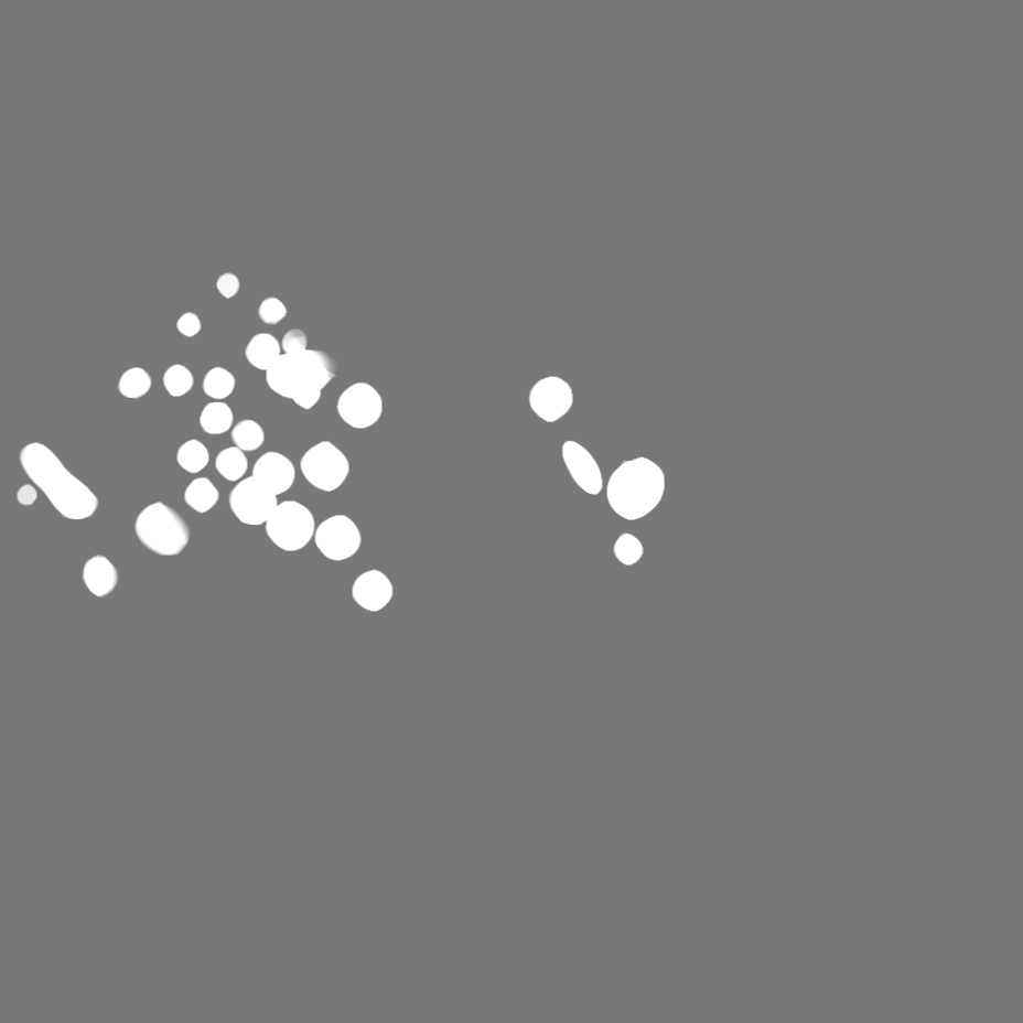
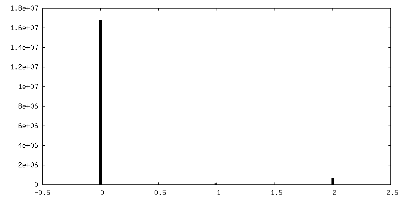
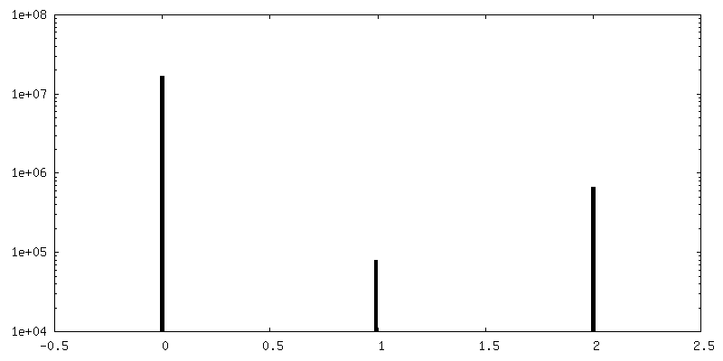
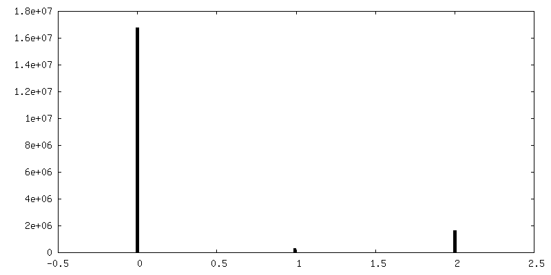
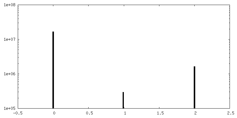
-Mask #1
| File | emd_10766_msk_1.map | ||||||||||||
|---|---|---|---|---|---|---|---|---|---|---|---|---|---|
| Projections & Slices |
| ||||||||||||
| Density Histograms |


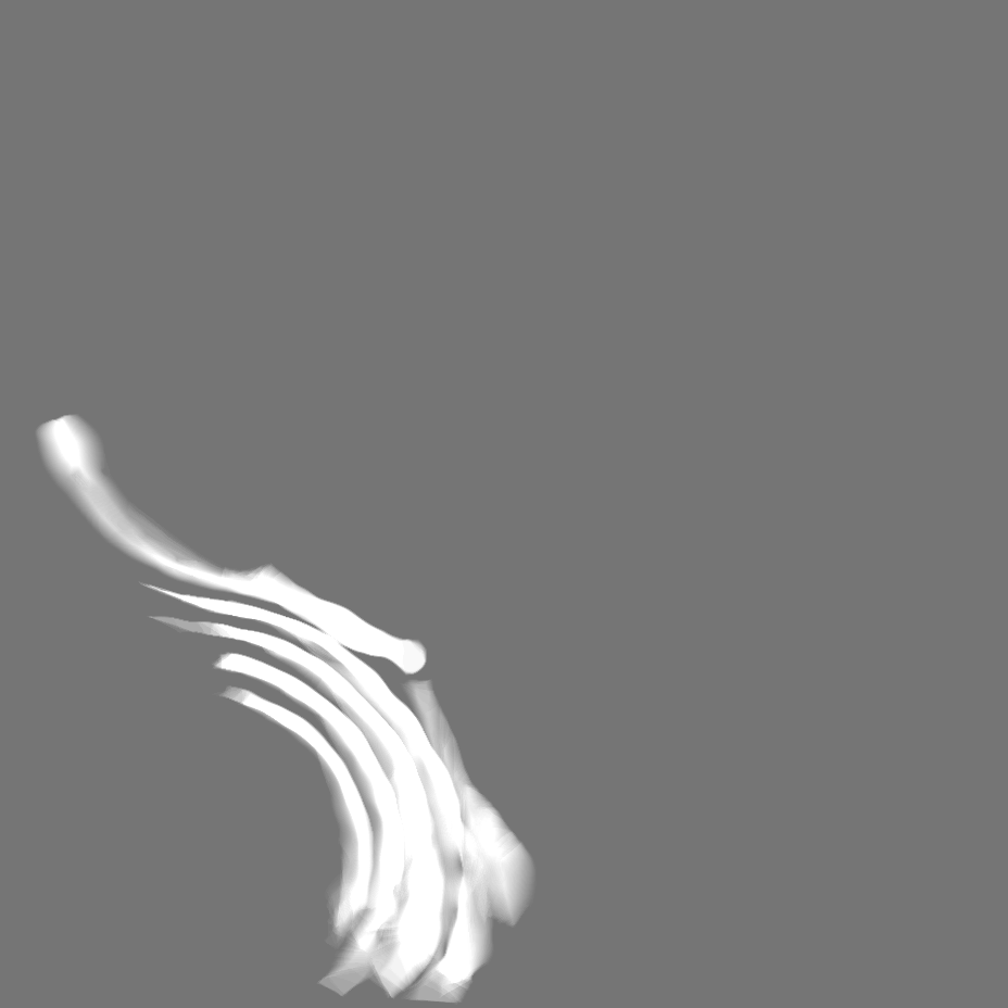
-Mask #2
| File | emd_10766_msk_2.map | ||||||||||||
|---|---|---|---|---|---|---|---|---|---|---|---|---|---|
| Projections & Slices |
| ||||||||||||
| Density Histograms |


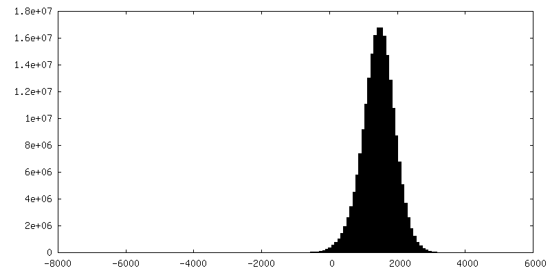
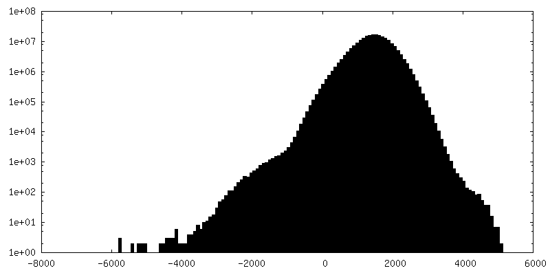
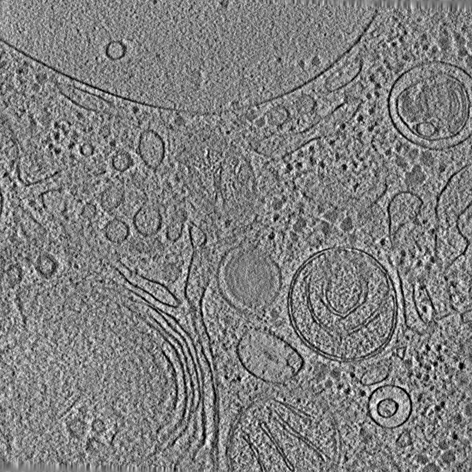
-Additional map: Filtered tomogram using a deconvolution filter (https://github.com/dtegunov/tom deconv) executed...
| File | emd_10766_additional.map | ||||||||||||
|---|---|---|---|---|---|---|---|---|---|---|---|---|---|
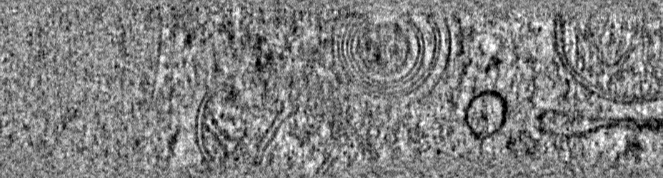
| Annotation | Filtered tomogram using a deconvolution filter (https://github.com/dtegunov/tom_deconv) executed in MATLAB (Mathworks) using the functionalities of the TOM toolbox (Nickel et al. 2005). | ||||||||||||
| Projections & Slices |
| ||||||||||||
| Density Histograms |


-Additional map: Filtered tomogram using a deconvolution filter (https://github.com/dtegunov/tom deconv) executed...
| File | emd_10766_additional_1.map | ||||||||||||
|---|---|---|---|---|---|---|---|---|---|---|---|---|---|
| Annotation | Filtered tomogram using a deconvolution filter (https://github.com/dtegunov/tom_deconv) executed in MATLAB (Mathworks) using the functionalities of the TOM toolbox (Nickel et al. 2005). | ||||||||||||
| Projections & Slices |
| ||||||||||||
| Density Histograms |


- Sample components
-Entire : Neurons from cortical mouse tissue cultured 7 days in vitro.
| Entire | Name: Neurons from cortical mouse tissue cultured 7 days in vitro. |
|---|---|
| Components |
|
-Supramolecule #1: Neurons from cortical mouse tissue cultured 7 days in vitro.
| Supramolecule | Name: Neurons from cortical mouse tissue cultured 7 days in vitro. type: organelle_or_cellular_component / ID: 1 / Parent: 0 |
|---|---|
| Source (natural) | Organism: |
| Recombinant expression | Organism: |
-Experimental details
-Structure determination
| Method | cryo EM |
|---|---|
 Processing Processing | electron tomography |
| Aggregation state | cell |
-Sample preparation
| Buffer | pH: 6 |
|---|---|
| Vitrification | Cryogen name: ETHANE-PROPANE |
| Sectioning | Focused ion beam - Instrument: OTHER / Focused ion beam - Ion: OTHER / Focused ion beam - Voltage: 30 kV / Focused ion beam - Current: 0.03 nA / Focused ion beam - Duration: 4000 sec. / Focused ion beam - Temperature: 90 K / Focused ion beam - Initial thickness: 1000 nm / Focused ion beam - Final thickness: 200 nm Focused ion beam - Details: The value given for _emd_sectioning_focused_ion_beam.instrument is Quanta 3D Cryo-FIB / SEM. This is not in a list of allowed values {'DB235', 'OTHER'} so OTHER is written into the XML file. |
- Electron microscopy
| Microscope | FEI TITAN KRIOS |
|---|---|
| Temperature | Min: 80.0 K / Max: 90.0 K |
| Specialist optics | Phase plate: VOLTA PHASE PLATE / Energy filter - Name: GIF Quantum LS / Energy filter - Slit width: 20 eV |
| Details | Also phase plate alignment. |
| Image recording | Film or detector model: GATAN K2 SUMMIT (4k x 4k) / Detector mode: COUNTING / Average electron dose: 2.0 e/Å2 |
| Electron beam | Acceleration voltage: 300 kV / Electron source:  FIELD EMISSION GUN FIELD EMISSION GUN |
| Electron optics | C2 aperture diameter: 50.1 µm / Illumination mode: OTHER / Imaging mode: BRIGHT FIELD / Nominal magnification: 33000 |
| Sample stage | Specimen holder model: FEI TITAN KRIOS AUTOGRID HOLDER / Cooling holder cryogen: NITROGEN |
| Experimental equipment |  Model: Titan Krios / Image courtesy: FEI Company |
-Image processing
| Final reconstruction | Algorithm: BACK PROJECTION / Software - Name: IMOD / Number images used: 60 |
|---|
