Journal: Nature / Year: 2020 Title: Stress- and ubiquitylation-dependent phase separation of the proteasome. Authors: Sayaka Yasuda / Hikaru Tsuchiya / Ai Kaiho / Qiang Guo / Ken Ikeuchi / Akinori Endo / Naoko Arai / Fumiaki Ohtake / Shigeo Murata / Toshifumi Inada / Wolfgang Baumeister / Rubén Fernández- ...Authors: Sayaka Yasuda / Hikaru Tsuchiya / Ai Kaiho / Qiang Guo / Ken Ikeuchi / Akinori Endo / Naoko Arai / Fumiaki Ohtake / Shigeo Murata / Toshifumi Inada / Wolfgang Baumeister / Rubén Fernández-Busnadiego / Keiji Tanaka / Yasushi Saeki / Abstract: The proteasome is a major proteolytic machine that regulates cellular proteostasis through selective degradation of ubiquitylated proteins. A number of ubiquitin-related molecules have recently been ...The proteasome is a major proteolytic machine that regulates cellular proteostasis through selective degradation of ubiquitylated proteins. A number of ubiquitin-related molecules have recently been found to be involved in the regulation of biomolecular condensates or membraneless organelles, which arise by liquid-liquid phase separation of specific biomolecules, including stress granules, nuclear speckles and autophagosomes, but it remains unclear whether the proteasome also participates in such regulation. Here we reveal that proteasome-containing nuclear foci form under acute hyperosmotic stress. These foci are transient structures that contain ubiquitylated proteins, p97 (also known as valosin-containing protein (VCP)) and multiple proteasome-interacting proteins, which collectively constitute a proteolytic centre. The major substrates for degradation by these foci were ribosomal proteins that failed to properly assemble. Notably, the proteasome foci exhibited properties of liquid droplets. RAD23B, a substrate-shuttling factor for the proteasome, and ubiquitylated proteins were necessary for formation of proteasome foci. In mechanistic terms, a liquid-liquid phase separation was triggered by multivalent interactions of two ubiquitin-associated domains of RAD23B and ubiquitin chains consisting of four or more ubiquitin molecules. Collectively, our results suggest that ubiquitin-chain-dependent phase separation induces the formation of a nuclear proteolytic compartment that promotes proteasomal degradation.
A: 12667.681 Å / B: 12667.681 Å / C: 5130.0 Å α=β=γ: 90.0 °
CCP4 map header:
mode
Image stored as Reals
Å/pix. X/Y/Z
13.680001079914
13.680001079914
13.68
M x/y/z
926
926
375
origin x/y/z
0.000
0.000
0.000
length x/y/z
12667.681
12667.681
5130.000
α/β/γ
90.000
90.000
90.000
MAP C/R/S
1
2
3
start NC/NR/NS
0
0
0
NC/NR/NS
926
926
375
D min/max/mean
-18667.459
6336.026
-0.003
-
Supplemental data
-
Sample components
-
Entire : nuclear region of HCT116 cell, following 0.2M sucrose stimulation
Entire
Name: nuclear region of HCT116 cell, following 0.2M sucrose stimulation
Components
Cell: nuclear region of HCT116 cell, following 0.2M sucrose stimulation
-
Supramolecule #1: nuclear region of HCT116 cell, following 0.2M sucrose stimulation
Supramolecule
Name: nuclear region of HCT116 cell, following 0.2M sucrose stimulation type: cell / ID: 1 / Parent: 0
Source (natural)
Organism: Homo sapiens (human)
-
Experimental details
-
Structure determination
Method
cryo EM
Processing
electron tomography
Aggregation state
cell
-
Sample preparation
Buffer
pH: 7
Grid
Model: Quantifoil R2/1 / Material: GOLD / Mesh: 200 / Support film - Material: CARBON / Support film - topology: HOLEY ARRAY
Vitrification
Cryogen name: ETHANE-PROPANE / Instrument: FEI VITROBOT MARK IV
Sectioning
Focused ion beam - Instrument: OTHER / Focused ion beam - Ion: OTHER / Focused ion beam - Voltage: 30 kV / Focused ion beam - Current: 0.01 nA / Focused ion beam - Duration: 1 sec. / Focused ion beam - Temperature: 93 K / Focused ion beam - Initial thickness: 1000 nm / Focused ion beam - Final thickness: 200 nm Focused ion beam - Details: The value given for _emd_sectioning_focused_ion_beam.instrument is Quanta 3D FEG, FEI. This is not in a list of allowed values {'DB235', 'OTHER'} so OTHER is written into the XML file.
-
Electron microscopy
Microscope
FEI TITAN KRIOS
Specialist optics
Energy filter - Slit width: 20 eV
Image recording
Film or detector model: GATAN K2 SUMMIT (4k x 4k) / Detector mode: COUNTING / Average electron dose: 1.8 e/Å2
Electron beam
Acceleration voltage: 300 kV / Electron source: FIELD EMISSION GUN
In the structure databanks used in Yorodumi, some data are registered as the other names, "COVID-19 virus" and "2019-nCoV". Here are the details of the virus and the list of structure data.
Jan 31, 2019. EMDB accession codes are about to change! (news from PDBe EMDB page)
EMDB accession codes are about to change! (news from PDBe EMDB page)
The allocation of 4 digits for EMDB accession codes will soon come to an end. Whilst these codes will remain in use, new EMDB accession codes will include an additional digit and will expand incrementally as the available range of codes is exhausted. The current 4-digit format prefixed with “EMD-” (i.e. EMD-XXXX) will advance to a 5-digit format (i.e. EMD-XXXXX), and so on. It is currently estimated that the 4-digit codes will be depleted around Spring 2019, at which point the 5-digit format will come into force.
The EM Navigator/Yorodumi systems omit the EMD- prefix.
Related info.:Q: What is EMD? / ID/Accession-code notation in Yorodumi/EM Navigator
Yorodumi is a browser for structure data from EMDB, PDB, SASBDB, etc.
This page is also the successor to EM Navigator detail page, and also detail information page/front-end page for Omokage search.
The word "yorodu" (or yorozu) is an old Japanese word meaning "ten thousand". "mi" (miru) is to see.
Related info.:EMDB / PDB / SASBDB / Comparison of 3 databanks / Yorodumi Search / Aug 31, 2016. New EM Navigator & Yorodumi / Yorodumi Papers / Jmol/JSmol / Function and homology information / Changes in new EM Navigator and Yorodumi
 Movie
Movie Controller

 Yorodumi
 Open data
 Basic information
 Map data
 Sample
 Homo sapiens (human)
 Authors
 Japan,
  Germany, 2 items
  Citation
 Structure visualization
 Movie viewer
 Downloads & links
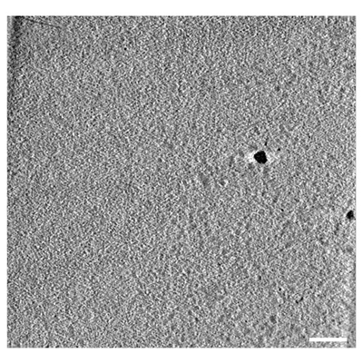 emd_10494.png
 http://ftp.pdbj.org/pub/emdb/structures/EMD-10494
 Z (Sec.)
 Y (Row.)
 X (Col.)


 Sample components
 Processing
 Electron microscopy
 FIELD EMISSION GUN
