United Kingdom, Switzerland, Germany, Spain, 11 items
Organization
Grant number
Country
Medical Research Council (United Kingdom)
MC_UU_00012/6
United Kingdom
Swiss National Science Foundation
31003A_179517
Switzerland
European Research Council
ERC-2012-SyG_318987-ToPAG
Germany
German Research Foundation
EXC 2067/1- 390729940
Germany
European Research Council
ERC AdG TENDO - 834394
Switzerland
Spanish Ministry of Economy and Competitiveness
SEV-2015-0522
Spain
German Research Foundation
SFB1190/P22
Germany
Spanish Ministry of Economy and Competitiveness
FIS2017-89560-R
Spain
Spanish Ministry of Economy and Competitiveness
FIS2015-63550-R
Spain
Spanish Ministry of Economy and Competitiveness
RYC-2017-22227
Spain
Spanish Ministry of Economy and Competitiveness
BFU2015-73288-JIN
Spain
Citation
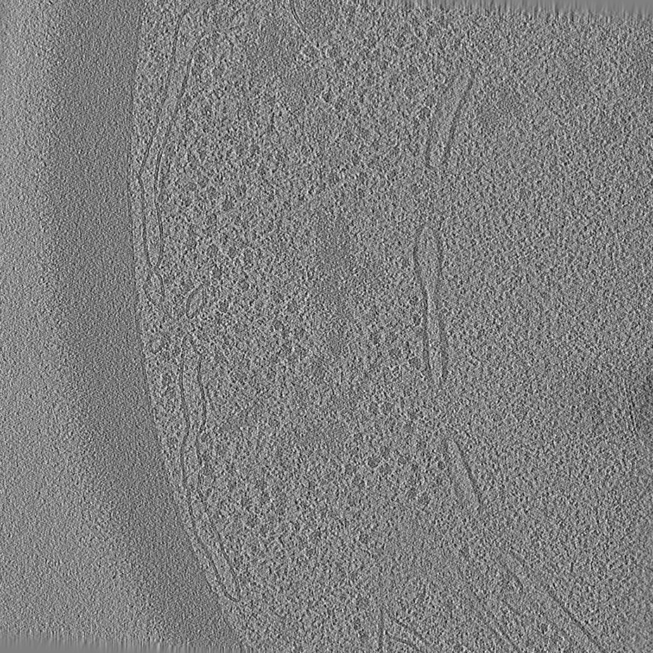
Journal: Dev Cell / Year: 2019 Title: Tricalbin-Mediated Contact Sites Control ER Curvature to Maintain Plasma Membrane Integrity. Authors: Javier Collado / Maria Kalemanov / Felix Campelo / Clélia Bourgoint / Ffion Thomas / Robbie Loewith / Antonio Martínez-Sánchez / Wolfgang Baumeister / Christopher J Stefan / Rubén Fernández-Busnadiego / Abstract: Membrane contact sites (MCS) between the endoplasmic reticulum (ER) and the plasma membrane (PM) play fundamental roles in all eukaryotic cells. ER-PM MCS are particularly abundant in Saccharomyces ...Membrane contact sites (MCS) between the endoplasmic reticulum (ER) and the plasma membrane (PM) play fundamental roles in all eukaryotic cells. ER-PM MCS are particularly abundant in Saccharomyces cerevisiae, where approximately half of the PM surface is covered by cortical ER (cER). Several proteins, including Ist2, Scs2/22, and Tcb1/2/3 are implicated in cER formation, but the specific roles of these molecules are poorly understood. Here, we use cryo-electron tomography to show that ER-PM tethers are key determinants of cER morphology. Notably, Tcb proteins (tricalbins) form peaks of extreme curvature on the cER membrane facing the PM. Combined modeling and functional assays suggest that Tcb-mediated cER peaks facilitate the transport of lipids between the cER and the PM, which is necessary to maintain PM integrity under heat stress. ER peaks were also present at other MCS, implying that membrane curvature enforcement may be a widespread mechanism to regulate MCS function.
Model: Quantifoil R2/1 / Material: COPPER / Mesh: 200 / Support film - Material: CARBON / Support film - topology: HOLEY / Pretreatment - Type: PLASMA CLEANING / Pretreatment - Atmosphere: AIR / Pretreatment - Pressure: 101.325 kPa
Vitrification
Cryogen name: ETHANE / Chamber humidity: 21 % / Chamber temperature: 300 K / Instrument: FEI VITROBOT MARK IV
Sectioning
Focused ion beam - Instrument: OTHER / Focused ion beam - Ion: OTHER / Focused ion beam - Voltage: 30 kV / Focused ion beam - Current: 0.03 nA / Focused ion beam - Duration: 7200 sec. / Focused ion beam - Temperature: 90 K / Focused ion beam - Initial thickness: 4000 nm / Focused ion beam - Final thickness: 200 nm Focused ion beam - Details: The value given for _emd_sectioning_focused_ion_beam.instrument is FEI Quanta FIB. This is not in a list of allowed values set(['DB235', 'OTHER']) so OTHER is written into the XML file.
-
Electron microscopy
Microscope
FEI TITAN KRIOS
Temperature
Min: 80.0 K / Max: 100.0 K
Specialist optics
Energy filter - Name: GIF Quantum LS / Energy filter - Slit width: 20 eV
Image recording
Film or detector model: GATAN K2 SUMMIT (4k x 4k) / Detector mode: COUNTING / Digitization - Dimensions - Width: 3838 pixel / Digitization - Dimensions - Height: 3710 pixel / Number grids imaged: 1 / Average exposure time: 1.8 sec. / Average electron dose: 1.7 e/Å2
Electron beam
Acceleration voltage: 300 kV / Electron source: FIELD EMISSION GUN
In the structure databanks used in Yorodumi, some data are registered as the other names, "COVID-19 virus" and "2019-nCoV". Here are the details of the virus and the list of structure data.
Jan 31, 2019. EMDB accession codes are about to change! (news from PDBe EMDB page)
EMDB accession codes are about to change! (news from PDBe EMDB page)
The allocation of 4 digits for EMDB accession codes will soon come to an end. Whilst these codes will remain in use, new EMDB accession codes will include an additional digit and will expand incrementally as the available range of codes is exhausted. The current 4-digit format prefixed with “EMD-” (i.e. EMD-XXXX) will advance to a 5-digit format (i.e. EMD-XXXXX), and so on. It is currently estimated that the 4-digit codes will be depleted around Spring 2019, at which point the 5-digit format will come into force.
The EM Navigator/Yorodumi systems omit the EMD- prefix.
Related info.:Q: What is EMD? / ID/Accession-code notation in Yorodumi/EM Navigator
Yorodumi is a browser for structure data from EMDB, PDB, SASBDB, etc.
This page is also the successor to EM Navigator detail page, and also detail information page/front-end page for Omokage search.
The word "yorodu" (or yorozu) is an old Japanese word meaning "ten thousand". "mi" (miru) is to see.
Related info.:EMDB / PDB / SASBDB / Comparison of 3 databanks / Yorodumi Search / Aug 31, 2016. New EM Navigator & Yorodumi / Yorodumi Papers / Jmol/JSmol / Function and homology information / Changes in new EM Navigator and Yorodumi
 Movie
Movie Controller

 Yorodumi
 Open data
 Basic information
 Map data
 Sample
 Authors
 United Kingdom,
  Switzerland,
  Germany,
  Spain, 11 items
  Citation
 Structure visualization
 Movie viewer
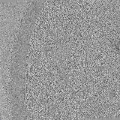
 Downloads & links
 emd_10378.png
 http://ftp.pdbj.org/pub/emdb/structures/EMD-10378
 Z (Sec.)
 Y (Row.)
 X (Col.)


 Sample components
 Processing
 Electron microscopy
 FIELD EMISSION GUN
