Journal: mBio / Year: 2020 Title: Diversification of Campylobacter jejuni Flagellar C-Ring Composition Impacts Its Structure and Function in Motility, Flagellar Assembly, and Cellular Processes. Authors: Louie D Henderson / Teige R S Matthews-Palmer / Connor J Gulbronson / Deborah A Ribardo / Morgan Beeby / David R Hendrixson / Abstract: Bacterial flagella are reversible rotary motors that rotate external filaments for bacterial propulsion. Some flagellar motors have diversified by recruiting additional components that influence ...Bacterial flagella are reversible rotary motors that rotate external filaments for bacterial propulsion. Some flagellar motors have diversified by recruiting additional components that influence torque and rotation, but little is known about the possible diversification and evolution of core motor components. The mechanistic core of flagella is the cytoplasmic C ring, which functions as a rotor, directional switch, and assembly platform for the flagellar type III secretion system (fT3SS) ATPase. The C ring is composed of a ring of FliG proteins and a helical ring of surface presentation of antigen (SPOA) domains from the switch proteins FliM and one of two usually mutually exclusive paralogs, FliN or FliY. We investigated the composition, architecture, and function of the C ring of , which encodes FliG, FliM, and both FliY and FliN by a variety of interrogative approaches. We discovered a diversified C ring containing FliG, FliM, and both FliY, which functions as a classical FliN-like protein for flagellar assembly, and FliN, which has neofunctionalized into a structural role. Specific protein interactions drive the formation of a more complex heterooligomeric C-ring structure. We discovered that this complex C ring has additional cellular functions in polarly localizing FlhG for numerical regulation of flagellar biogenesis and spatial regulation of division. Furthermore, mutation of the C ring revealed a T3SS that was less dependent on its ATPase complex for assembly than were other systems. Our results highlight considerable evolved flagellar diversity that impacts motor output, biogenesis, and cellular processes in different species. The conserved core of bacterial flagellar motors reflects a shared evolutionary history that preserves the mechanisms essential for flagellar assembly, rotation, and directional switching. In this work, we describe an expanded and diversified set of core components in the flagellar C ring, the mechanistic core of the motor. Our work provides insight into how usually conserved core components may have diversified by gene duplication, enabling a division of labor of the ancestral protein between the two new proteins, acquisition of new roles in flagellar assembly and motility, and expansion of the function of the flagellum beyond motility, including spatial regulation of cell division and numerical control of flagellar biogenesis in Our results highlight that relatively small changes, such as gene duplications, can have substantial ramifications on the cellular roles of a molecular machine.
Entire : Bacterial flagellar motor (cellular-component from Campylobacter ...
Entire
Name: Bacterial flagellar motor (cellular-component from Campylobacter jejuni, located in [Cell wall])
Components
Organelle or cellular component: Bacterial flagellar motor (cellular-component from Campylobacter jejuni, located in [Cell wall])
-
Supramolecule #1: Bacterial flagellar motor (cellular-component from Campylobacter ...
Supramolecule
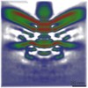
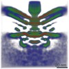
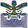
Name: Bacterial flagellar motor (cellular-component from Campylobacter jejuni, located in [Cell wall]) type: organelle_or_cellular_component / ID: 1 / Parent: 0 / Macromolecule list: #1
Source (natural)
Organism: Campylobacter jejuni subsp. jejuni 81-176 (Campylobacter) Location in cell: Cell pole
-
Experimental details
-
Structure determination
Method
cryo EM
Processing
subtomogram averaging
Aggregation state
cell
-
Sample preparation
Buffer
pH: 7
Vitrification
Cryogen name: ETHANE-PROPANE / Chamber humidity: 95 % / Chamber temperature: 298 K / Instrument: FEI VITROBOT MARK IV / Details: Blot time 2, blot force 2, OD 20.
Details
In situ within Campylobacter jejuni
-
Electron microscopy
Microscope
FEI TECNAI F20
Image recording
Film or detector model: FEI FALCON II (4k x 4k) / Average electron dose: 120.0 e/Å2
Electron beam
Acceleration voltage: 200 kV / Electron source: FIELD EMISSION GUN
In the structure databanks used in Yorodumi, some data are registered as the other names, "COVID-19 virus" and "2019-nCoV". Here are the details of the virus and the list of structure data.
Jan 31, 2019. EMDB accession codes are about to change! (news from PDBe EMDB page)
EMDB accession codes are about to change! (news from PDBe EMDB page)
The allocation of 4 digits for EMDB accession codes will soon come to an end. Whilst these codes will remain in use, new EMDB accession codes will include an additional digit and will expand incrementally as the available range of codes is exhausted. The current 4-digit format prefixed with “EMD-” (i.e. EMD-XXXX) will advance to a 5-digit format (i.e. EMD-XXXXX), and so on. It is currently estimated that the 4-digit codes will be depleted around Spring 2019, at which point the 5-digit format will come into force.
The EM Navigator/Yorodumi systems omit the EMD- prefix.
Related info.:Q: What is EMD? / ID/Accession-code notation in Yorodumi/EM Navigator
Yorodumi is a browser for structure data from EMDB, PDB, SASBDB, etc.
This page is also the successor to EM Navigator detail page, and also detail information page/front-end page for Omokage search.
The word "yorodu" (or yorozu) is an old Japanese word meaning "ten thousand". "mi" (miru) is to see.
Related info.:EMDB / PDB / SASBDB / Comparison of 3 databanks / Yorodumi Search / Aug 31, 2016. New EM Navigator & Yorodumi / Yorodumi Papers / Jmol/JSmol / Function and homology information / Changes in new EM Navigator and Yorodumi
 Movie
Movie Controller


 Open data
 Basic information
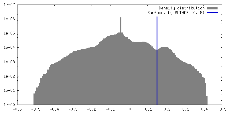
 Map data
 Sample
 Campylobacter jejuni subsp. jejuni 81-176 (Campylobacter)
 Authors
 United Kingdom, 1 items
  Citation
 Structure visualization
 Movie viewer
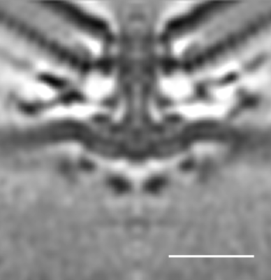
 Downloads & links
 emd_10343.png
 http://ftp.pdbj.org/pub/emdb/structures/EMD-10343

 Z (Sec.)
 Y (Row.)
 X (Col.)


 Sample components
 Processing
 Electron microscopy
 FIELD EMISSION GUN
