 Movie
Movie Controller

[English] 日本語
 Yorodumi
- EMDB-10309: cryo-ET of cryo-FIB milled yeast cell, in which scs2/22 ist2 are ... -
+ Open data

- Basic information
| Entry | Database: EMDB / ID: EMD-10309 | ||||||||||||
|---|---|---|---|---|---|---|---|---|---|---|---|---|---|
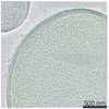
| Title | cryo-ET of cryo-FIB milled yeast cell, in which scs2/22 ist2 are deleted, with high intracellular calcium; shown in Figures 5C and S3F | ||||||||||||
 Map data Map data | cryo-ET of cryo-FIB milled yeast cell in which scs2/22 ist2 are deleted with high calcium levels; shown in Figures 5C and S3F | ||||||||||||
 Sample Sample |
| ||||||||||||
| Biological species |  | ||||||||||||
| Method | electron tomography / cryo EM | ||||||||||||
 Authors Authors | Hoffmann PC / Bharat TAM / Wozny MR / Boulanger J / Miller EA / Kukulski W | ||||||||||||
| Funding support |  United Kingdom, 3 items United Kingdom, 3 items
| ||||||||||||
 Citation Citation | Journal: Dev Cell / Year: 2019 Title: Tricalbins Contribute to Cellular Lipid Flux and Form Curved ER-PM Contacts that Are Bridged by Rod-Shaped Structures. Authors: Patrick C Hoffmann / Tanmay A M Bharat / Michael R Wozny / Jerome Boulanger / Elizabeth A Miller / Wanda Kukulski / Abstract: Lipid flow between cellular organelles occurs via membrane contact sites. Extended-synaptotagmins, known as tricalbins in yeast, mediate lipid transfer between the endoplasmic reticulum (ER) and ...Lipid flow between cellular organelles occurs via membrane contact sites. Extended-synaptotagmins, known as tricalbins in yeast, mediate lipid transfer between the endoplasmic reticulum (ER) and plasma membrane (PM). How these proteins regulate membrane architecture to transport lipids across the aqueous space between bilayers remains unknown. Using correlative microscopy, electron cryo-tomography, and high-throughput genetics, we address the interplay of architecture and function in budding yeast. We find that ER-PM contacts differ in protein composition and membrane morphology, not in intermembrane distance. In situ electron cryo-tomography reveals the molecular organization of tricalbin-mediated contacts, suggesting a structural framework for putative lipid transfer. Genetic analysis uncovers functional overlap with cellular lipid routes, such as maintenance of PM asymmetry. Further redundancies are suggested for individual tricalbin protein domains. We propose a modularity of molecular and structural functions of tricalbins and of their roles within the cellular network of lipid distribution pathways. | ||||||||||||
| History |
|
- Structure visualization
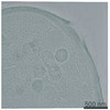
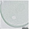
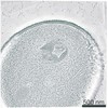
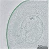
| Movie |
 Movie viewer Movie viewer |
|---|---|
| Supplemental images |

- Downloads & links
-EMDB archive
| Map data | emd_10309.map.gz | 1.1 GB | EMDB map data format | |
|---|---|---|---|---|
| Header (meta data) | emd-10309-v30.xmlemd-10309.xml | 13.2 KB 13.2 KB | Display Display | EMDB header |
| Images | 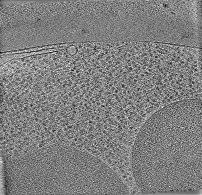 emd_10309.png emd_10309.png | 140.3 KB | ||
| Archive directory |  http://ftp.pdbj.org/pub/emdb/structures/EMD-10309ftp://ftp.pdbj.org/pub/emdb/structures/EMD-10309 http://ftp.pdbj.org/pub/emdb/structures/EMD-10309ftp://ftp.pdbj.org/pub/emdb/structures/EMD-10309 | HTTPS FTP |
-Related structure data
| Related structure data | C: citing same article ( |
|---|---|
| EM raw data | EMPIAR-10321 (Title: cryo-ET of cryo-FIB milled yeast cell in which scs2/22 ist2 are deleted with high intracellular calcium Data size: 14.3 Data #1: cryo-ET of cryo-FIB milled yeast cell with high intracellular calcium (scs2/22 ist2 deletion and GCaMP expression) [tilt series]) |
-Links
| EMDB pages | EMDB (EBI/PDBe) / EMDataResource |
|---|
-Map
| File | Download / File: emd_10309.map.gz / Format: CCP4 / Size: 1.4 GB / Type: IMAGE STORED AS SIGNED BYTE | ||||||||||||||||||||||||||||||||||||||||||||||||||||||||||||||||||||
|---|---|---|---|---|---|---|---|---|---|---|---|---|---|---|---|---|---|---|---|---|---|---|---|---|---|---|---|---|---|---|---|---|---|---|---|---|---|---|---|---|---|---|---|---|---|---|---|---|---|---|---|---|---|---|---|---|---|---|---|---|---|---|---|---|---|---|---|---|---|
| Annotation | cryo-ET of cryo-FIB milled yeast cell in which scs2/22 ist2 are deleted with high calcium levels; shown in Figures 5C and S3F | ||||||||||||||||||||||||||||||||||||||||||||||||||||||||||||||||||||
| Voxel size | X=Y=Z: 7.4 Å | ||||||||||||||||||||||||||||||||||||||||||||||||||||||||||||||||||||
| Density |
| ||||||||||||||||||||||||||||||||||||||||||||||||||||||||||||||||||||
| Symmetry | Space group: 1 | ||||||||||||||||||||||||||||||||||||||||||||||||||||||||||||||||||||
| Details | EMDB XML:
CCP4 map header:
| ||||||||||||||||||||||||||||||||||||||||||||||||||||||||||||||||||||
-Supplemental data
- Sample components
-Entire : cryo-ET of cryo-FIB milled yeast cell, in which scs2/22 ist2 are ...
| Entire | Name: cryo-ET of cryo-FIB milled yeast cell, in which scs2/22 ist2 are deleted, with high intracellular calcium; shown in Figures 5C and S3F |
|---|---|
| Components |
|
-Supramolecule #1: cryo-ET of cryo-FIB milled yeast cell, in which scs2/22 ist2 are ...
| Supramolecule | Name: cryo-ET of cryo-FIB milled yeast cell, in which scs2/22 ist2 are deleted, with high intracellular calcium; shown in Figures 5C and S3F type: cell / ID: 1 / Parent: 0 |
|---|---|
| Source (natural) | Organism: |
-Experimental details
-Structure determination
| Method | cryo EM |
|---|---|
 Processing Processing | electron tomography |
| Aggregation state | cell |
-Sample preparation
| Buffer | pH: 5.5 Details: Synthetic Complete -Trp medium with 15% high molecular weight dextran (w/v), 2 % glucose and 200 mM calcium chloride |
|---|---|
| Vitrification | Cryogen name: ETHANE / Chamber temperature: 298 K / Instrument: HOMEMADE PLUNGER Details: 5 microliter of cell suspension was applied to the grid and then backside-blotted for 12-15 s. |
| Details | Cells were treated with 200 mM calcium chloride before plunge freezing. The cells with high GCaMP fluorescent signals, indicating high calcium levels, were targeted for cryo-FIB milling |
| Cryo protectant | dextran |
| Sectioning | Focused ion beam - Instrument: OTHER / Focused ion beam - Ion: OTHER / Focused ion beam - Voltage: 16 kV / Focused ion beam - Current: 0.023 nA / Focused ion beam - Duration: 3600 sec. / Focused ion beam - Temperature: 103 K / Focused ion beam - Initial thickness: 5000 nm / Focused ion beam - Final thickness: 200 nm Focused ion beam - Details: Initial rough milling was performed at 30 kV 0.5-1 nA, then with 0.3 nA until 3000 nm thickness, then decreased to 0.1 nA to 1000 nm thickness. Fine milling to 150-300 nm ...Focused ion beam - Details: Initial rough milling was performed at 30 kV 0.5-1 nA, then with 0.3 nA until 3000 nm thickness, then decreased to 0.1 nA to 1000 nm thickness. Fine milling to 150-300 nm thickness was performed at 16kV 11 pA or 23 pA.. The value given for _emd_sectioning_focused_ion_beam.instrument is Scios DualBeam. This is not in a list of allowed values set(['DB235', 'OTHER']) so OTHER is written into the XML file. |
- Electron microscopy
| Microscope | FEI TITAN KRIOS |
|---|---|
| Details | Montaged images of the grid were acquired at 200 nm pixel size to localize the lamellae on the grid. Overview montages of the individual lamellae were acquired at about 5 nm pixel size to assess the lamellae quality and identify ER-PM contact sites within the cells |
| Image recording | Film or detector model: GATAN K2 SUMMIT (4k x 4k) / Detector mode: COUNTING / Digitization - Frames/image: 1-4 / Average electron dose: 1.0 e/Å2 Details: Electron cryo-tomographic tilt-series were collected on a Titan Krios (FEI) operated at 300 kV using a Quantum energy filter (slit width 20 eV) and a K2 direct electron detector (Gatan) in ...Details: Electron cryo-tomographic tilt-series were collected on a Titan Krios (FEI) operated at 300 kV using a Quantum energy filter (slit width 20 eV) and a K2 direct electron detector (Gatan) in counting mode at a pixel size of 3.7 angstroms and at a dose rate of ~ 2-4 e-/pixel/second on the detector. Tilt-series were acquired between +/- 60 degrees starting from 0 degrees with 1 degrees increment using SerialEM (Mastronarde, 2005) following a grouped dose-symmetric acquisition with a group size of 4 (Bharat et al., 2018; Hagen et al., 2017), and at -5 micron defocus. A dose of 1.0 e-/square angstroms was applied per image of the tilt-series. |
| Electron beam | Acceleration voltage: 300 kV / Electron source:  FIELD EMISSION GUN FIELD EMISSION GUN |
| Electron optics | Illumination mode: FLOOD BEAM / Imaging mode: BRIGHT FIELD |
| Experimental equipment |  Model: Titan Krios / Image courtesy: FEI Company |
-Image processing
| Final reconstruction | Algorithm: SIMULTANEOUS ITERATIVE (SIRT) / Software - Name: eTomo / Details: 10 SIRT iterations / Number images used: 106 |
|---|---|
| CTF correction | Software - Name: eTomo |
