 Movie
Movie Controller


+ Open data

- Basic information
| Entry | Database: EMDB / ID: EMD-0965 | |||||||||||||||
|---|---|---|---|---|---|---|---|---|---|---|---|---|---|---|---|---|
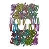
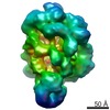
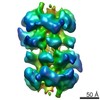
| Title | AAA+ ATPase, ClpL from Streptococcus pneumoniae - ATP bound | |||||||||||||||
 Map data Map data | ||||||||||||||||
 Sample Sample |
| |||||||||||||||
 Keywords Keywords | AAA+ ATPase / Chaperone / Streptococcus pneumoniae | |||||||||||||||
| Function / homology |  Function and homology information Function and homology informationpeptidase activity / cellular response to heat / ATP hydrolysis activity / proteolysis / ATP binding / cytoplasm Similarity search - Function | |||||||||||||||
| Biological species |   Streptococcus pneumoniae (bacteria) Streptococcus pneumoniae (bacteria) | |||||||||||||||
| Method | single particle reconstruction / cryo EM / Resolution: 6.33 Å | |||||||||||||||
 Authors Authors | Kim G / Lee SG | |||||||||||||||
| Funding support |  Korea, Republic Of, 4 items Korea, Republic Of, 4 items
| |||||||||||||||
 Citation Citation | Journal: FASEB J / Year: 2020 Title: ClpL is a functionally active tetradecameric AAA+ chaperone, distinct from hexameric/dodecameric ones. Authors: Gyuhee Kim / Seong-Gyu Lee / Seungsu Han / Jaeeun Jung / Hyeong Seop Jeong / Jae-Kyung Hyun / Dong-Kwon Rhee / Ho Min Kim / Sangho Lee /  Abstract: AAA+ (ATPases associated with diverse cellular activities) chaperones are involved in a plethora of cellular activities to ensure protein homeostasis. The function of AAA+ chaperones is mostly ...AAA+ (ATPases associated with diverse cellular activities) chaperones are involved in a plethora of cellular activities to ensure protein homeostasis. The function of AAA+ chaperones is mostly modulated by their hexameric/dodecameric quaternary structures. Here we report the structural and biochemical characterizations of a tetradecameric AAA+ chaperone, ClpL from Streptococcus pneumoniae. ClpL exists as a tetradecamer in solution in the presence of ATP. The cryo-EM structure of ClpL at 4.5 Å resolution reveals a striking tetradecameric arrangement. Solution structures of ClpL derived from small-angle X-ray scattering data suggest that the tetradecameric ClpL could assume a spiral conformation found in active hexameric/dodecameric AAA+ chaperone structures. Vertical positioning of the middle domain accounts for the head-to-head arrangement of two heptameric rings. Biochemical activity assays with site-directed mutagenesis confirmed the critical roles of residues both in the integrity of the tetradecameric arrangement and activities of ClpL. Non-conserved Q321 and R670 are crucial in the heptameric ring assembly of ClpL. These results establish that ClpL is a functionally active tetradecamer, clearly distinct from hexameric/dodecameric AAA+ chaperones. | |||||||||||||||
| History |
|
- Structure visualization
| Movie |
Movie viewer |
|---|---|
| Structure viewer | EM map: SurfViewMolmilJmol/JSmol |
| Supplemental images |

- Downloads & links
-EMDB archive
| Map data | emd_0965.map.gz | 6.4 MB | EMDB map data format | |
|---|---|---|---|---|
| Header (meta data) | emd-0965-v30.xmlemd-0965.xml | 12.9 KB 12.9 KB | Display Display | EMDB header |
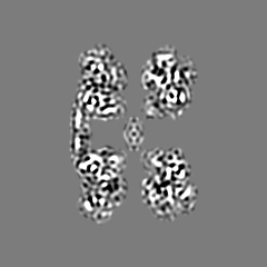
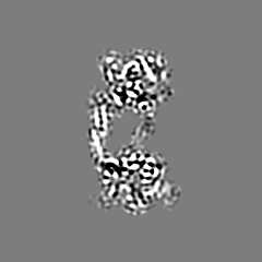
| Images |  emd_0965.png emd_0965.png | 140.2 KB | ||
| Filedesc metadata | emd-0965.cif.gz | 5.7 KB | ||
| Archive directory |  http://ftp.pdbj.org/pub/emdb/structures/EMD-0965ftp://ftp.pdbj.org/pub/emdb/structures/EMD-0965 http://ftp.pdbj.org/pub/emdb/structures/EMD-0965ftp://ftp.pdbj.org/pub/emdb/structures/EMD-0965 | HTTPS FTP |
-Related structure data
| Related structure data |  6lsyMC  0967C  6lt4C M: atomic model generated by this map C: citing same article ( |
|---|---|
| Similar structure data |

-Links
| EMDB pages | EMDB (EBI/PDBe) / EMDataResource |
|---|---|
| Related items in Molecule of the Month |


-Map
| File | Download / File: emd_0965.map.gz / Format: CCP4 / Size: 52.7 MB / Type: IMAGE STORED AS FLOATING POINT NUMBER (4 BYTES) | ||||||||||||||||||||||||||||||||||||||||||||||||||||||||||||
|---|---|---|---|---|---|---|---|---|---|---|---|---|---|---|---|---|---|---|---|---|---|---|---|---|---|---|---|---|---|---|---|---|---|---|---|---|---|---|---|---|---|---|---|---|---|---|---|---|---|---|---|---|---|---|---|---|---|---|---|---|---|
| Projections & slices | Image control
Images are generated by Spider. | ||||||||||||||||||||||||||||||||||||||||||||||||||||||||||||
| Voxel size | X=Y=Z: 1.4 Å | ||||||||||||||||||||||||||||||||||||||||||||||||||||||||||||
| Density |
| ||||||||||||||||||||||||||||||||||||||||||||||||||||||||||||
| Symmetry | Space group: 1 | ||||||||||||||||||||||||||||||||||||||||||||||||||||||||||||
| Details | EMDB XML:
CCP4 map header:
| ||||||||||||||||||||||||||||||||||||||||||||||||||||||||||||
 Z (Sec.)
 Y (Row.)
 X (Col.)


-Supplemental data
- Sample components
-Entire : ClpL Trap(E193A/E526A):ATP-bound
| Entire | Name: ClpL Trap(E193A/E526A):ATP-bound |
|---|---|
| Components |
|
-Supramolecule #1: ClpL Trap(E193A/E526A):ATP-bound
| Supramolecule | Name: ClpL Trap(E193A/E526A):ATP-bound / type: organelle_or_cellular_component / ID: 1 / Parent: 0 / Macromolecule list: all |
|---|---|
| Source (natural) | Organism: Streptococcus pneumoniae (bacteria) |
| Molecular weight | Theoretical: 1.1 MDa |
-Macromolecule #1: ATP-dependent Clp protease, ATP-binding subunit
| Macromolecule | Name: ATP-dependent Clp protease, ATP-binding subunit / type: protein_or_peptide / ID: 1 / Number of copies: 14 / Enantiomer: LEVO |
|---|---|
| Source (natural) | Organism: Streptococcus pneumoniae (bacteria) |
| Molecular weight | Theoretical: 77.599094 KDa |
| Recombinant expression | Organism: |
| Sequence | String: MNNNFNNFNN MDDLFNQLMG GMRGYSSENR RYLINGREVT PEEFAYYRAT GQLPGNAESD VQMQQQASGM KQDGVLAKLG RNLTAEARE GKLDPVIGRN KEIQEASEIL SRRTKNNPVL VGDAGVGKTA VVEGLAQAIV NGDVPAAIKN KEIVSIDISG L EAGTQYRG ...String: MNNNFNNFNN MDDLFNQLMG GMRGYSSENR RYLINGREVT PEEFAYYRAT GQLPGNAESD VQMQQQASGM KQDGVLAKLG RNLTAEARE GKLDPVIGRN KEIQEASEIL SRRTKNNPVL VGDAGVGKTA VVEGLAQAIV NGDVPAAIKN KEIVSIDISG L EAGTQYRG SFEENVQNLV NEVKEAGNII LFFDAIHQIL GAGSTGGDSG SKGLADILKP ALSRGELTVI GATTQDEYRN TI LKNAALA RRFNEVKVNA PSAENTFKIL QGIRDLYQQH HNVILPDEVL KAAVDYSVQY IPQRSLPDKA IDLVDVTAAH LAA QHPVTD VHAVEREIET EKDKQEKAVE AEDFEAALNY KTRIAELERK IENHTEDMKV TASVNDVAES VERMTGIPVS QMGA SDIER LKDMAHRLQD KVIGQDKAVE VVARAICRNR AGFDEGNRPI GNFLFVGSTG VGKTELAKQL ALDMFGTQDA IIRLD MSEY SDRTAVSKLI GTTAGYVGYD DNSNTLTERV RRNPYSIILL DAIEKADPQV ITLLLQVLDD GRLTDGQGNT VNFKNT VII ATSNAGFGYE ANLTEDADKP ELMDRLKPFF RPEFLNRFNA VIEFSHLTKE DLSKIVDLML AEVNQTLAKK DIDLVVS QA AKDYITEEGY DEVMGVRPLR RVVEQEIRDK VTDFHLDHLD AKHLEADMED GVLVIREKV UniProtKB: ATP-dependent Clp protease, ATP-binding subunit |
-Experimental details
-Structure determination
| Method | cryo EM |
|---|---|
 Processing Processing | single particle reconstruction |
| Aggregation state | particle |
-Sample preparation
| Concentration | 0.5 mg/mL | |||||||||||||||
|---|---|---|---|---|---|---|---|---|---|---|---|---|---|---|---|---|
| Buffer | pH: 7.5 Component:
| |||||||||||||||
| Grid | Model: Quantifoil R1.2/1.3 / Material: COPPER / Mesh: 300 | |||||||||||||||
| Vitrification | Cryogen name: ETHANE / Chamber humidity: 100 % / Chamber temperature: 277 K / Instrument: FEI VITROBOT MARK IV |
- Electron microscopy
| Microscope | FEI TITAN KRIOS |
|---|---|
| Image recording | Film or detector model: FEI FALCON III (4k x 4k) / Average electron dose: 60.0 e/Å2 |
| Electron beam | Acceleration voltage: 300 kV / Electron source:  FIELD EMISSION GUN FIELD EMISSION GUN |
| Electron optics | Illumination mode: FLOOD BEAM / Imaging mode: DARK FIELD |
| Experimental equipment |  Model: Titan Krios / Image courtesy: FEI Company |
+Image processing

-Atomic model buiding 1
| Refinement | Protocol: AB INITIO MODEL |
|---|---|
| Output model | PDB-6lsy: |
