Journal: Nature / Year: 2020 Title: Isolation of an archaeon at the prokaryote-eukaryote interface. Authors: Hiroyuki Imachi / Masaru K Nobu / Nozomi Nakahara / Yuki Morono / Miyuki Ogawara / Yoshihiro Takaki / Yoshinori Takano / Katsuyuki Uematsu / Tetsuro Ikuta / Motoo Ito / Yohei Matsui / ...Authors: Hiroyuki Imachi / Masaru K Nobu / Nozomi Nakahara / Yuki Morono / Miyuki Ogawara / Yoshihiro Takaki / Yoshinori Takano / Katsuyuki Uematsu / Tetsuro Ikuta / Motoo Ito / Yohei Matsui / Masayuki Miyazaki / Kazuyoshi Murata / Yumi Saito / Sanae Sakai / Chihong Song / Eiji Tasumi / Yuko Yamanaka / Takashi Yamaguchi / Yoichi Kamagata / Hideyuki Tamaki / Ken Takai / Abstract: The origin of eukaryotes remains unclear. Current data suggest that eukaryotes may have emerged from an archaeal lineage known as 'Asgard' archaea. Despite the eukaryote-like genomic features that ...The origin of eukaryotes remains unclear. Current data suggest that eukaryotes may have emerged from an archaeal lineage known as 'Asgard' archaea. Despite the eukaryote-like genomic features that are found in these archaea, the evolutionary transition from archaea to eukaryotes remains unclear, owing to the lack of cultured representatives and corresponding physiological insights. Here we report the decade-long isolation of an Asgard archaeon related to Lokiarchaeota from deep marine sediment. The archaeon-'Candidatus Prometheoarchaeum syntrophicum' strain MK-D1-is an anaerobic, extremely slow-growing, small coccus (around 550 nm in diameter) that degrades amino acids through syntrophy. Although eukaryote-like intracellular complexes have been proposed for Asgard archaea, the isolate has no visible organelle-like structure. Instead, Ca. P. syntrophicum is morphologically complex and has unique protrusions that are long and often branching. On the basis of the available data obtained from cultivation and genomics, and reasoned interpretations of the existing literature, we propose a hypothetical model for eukaryogenesis, termed the entangle-engulf-endogenize (also known as E) model.
Cryogen name: ETHANE / Chamber humidity: 95 % / Chamber temperature: 277 K / Instrument: FEI VITROBOT MARK IV
Sectioning
Other: NO SECTIONING
Fiducial marker
Manufacturer: Sigma-Aldrich / Diameter: 15 nm
-
Electron microscopy
Microscope
JEOL 2200FS
Temperature
Min: 76.0 K / Max: 77.0 K
Specialist optics
Energy filter - Name: In-column Omega Filter / Energy filter - Slit width: 20 eV
Image recording
Film or detector model: DIRECT ELECTRON DE-20 (5k x 3k) / Digitization - Dimensions - Width: 5120 pixel / Digitization - Dimensions - Height: 3840 pixel / Digitization - Frames/image: 3-25 / Number grids imaged: 1 / Number real images: 63 / Average exposure time: 1.0 sec. / Average electron dose: 1.5 e/Å2
Electron beam
Acceleration voltage: 200 kV / Electron source: FIELD EMISSION GUN
In the structure databanks used in Yorodumi, some data are registered as the other names, "COVID-19 virus" and "2019-nCoV". Here are the details of the virus and the list of structure data.
Jan 31, 2019. EMDB accession codes are about to change! (news from PDBe EMDB page)
EMDB accession codes are about to change! (news from PDBe EMDB page)
The allocation of 4 digits for EMDB accession codes will soon come to an end. Whilst these codes will remain in use, new EMDB accession codes will include an additional digit and will expand incrementally as the available range of codes is exhausted. The current 4-digit format prefixed with “EMD-” (i.e. EMD-XXXX) will advance to a 5-digit format (i.e. EMD-XXXXX), and so on. It is currently estimated that the 4-digit codes will be depleted around Spring 2019, at which point the 5-digit format will come into force.
The EM Navigator/Yorodumi systems omit the EMD- prefix.
Related info.:Q: What is EMD? / ID/Accession-code notation in Yorodumi/EM Navigator
Yorodumi is a browser for structure data from EMDB, PDB, SASBDB, etc.
This page is also the successor to EM Navigator detail page, and also detail information page/front-end page for Omokage search.
The word "yorodu" (or yorozu) is an old Japanese word meaning "ten thousand". "mi" (miru) is to see.
Related info.:EMDB / PDB / SASBDB / Comparison of 3 databanks / Yorodumi Search / Aug 31, 2016. New EM Navigator & Yorodumi / Yorodumi Papers / Jmol/JSmol / Function and homology information / Changes in new EM Navigator and Yorodumi
 Movie
Movie Controller

 Yorodumi
 Open data
 Basic information
 Map data
 Sample
 anaerobic archaeon MK-D1 (archaea)
 Authors
 Japan, 11 items
  Citation
 Structure visualization
 Movie viewer
 Downloads & links
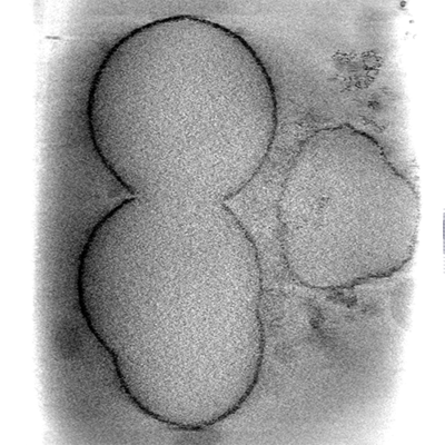 emd_0852.png
 http://ftp.pdbj.org/pub/emdb/structures/EMD-0852
 Sample components
 Processing
 Electron microscopy
 FIELD EMISSION GUN