 ムービー
ムービー コントローラー


+ データを開く

- 基本情報
基本情報
| 登録情報 | データベース: EMDB / ID: EMD-0733 | |||||||||
|---|---|---|---|---|---|---|---|---|---|---|
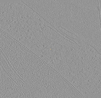
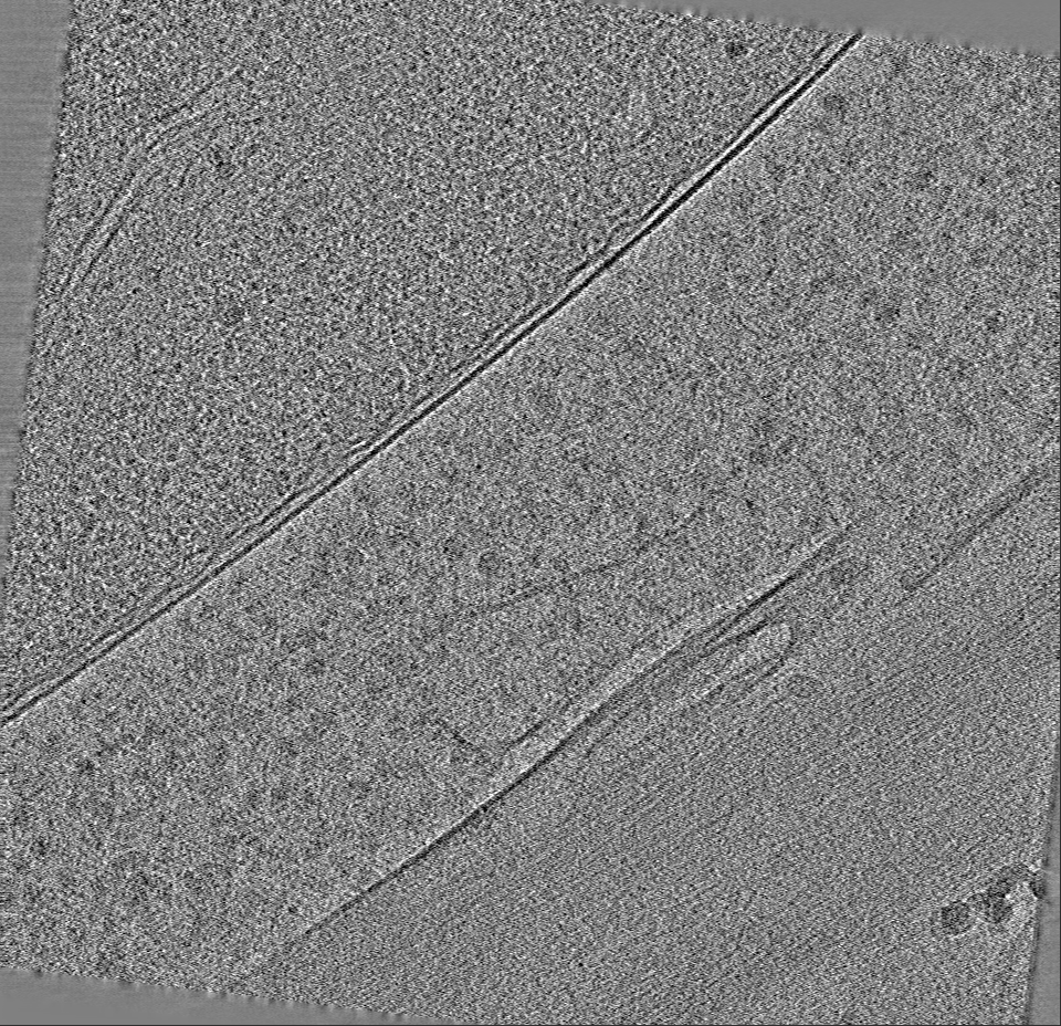
| タイトル | Cryo electron tomogram of cryo-lamella of spinach leaf | |||||||||
 マップデータ マップデータ | The pixel size of original image is 2.65, this reconstructed tomogram has a binning factor of 4. | |||||||||
 試料 試料 |
| |||||||||
| 生物種 |  Spinacia oleracea (ホウレンソウ) Spinacia oleracea (ホウレンソウ) | |||||||||
| 手法 | 電子線トモグラフィー法 / クライオ電子顕微鏡法 | |||||||||
 データ登録者 データ登録者 | Zhang J / Zhang D / Sun L / Sun F | |||||||||
| 資金援助 |  中国, 2件 中国, 2件
| |||||||||
 引用 引用 | ジャーナル: J Struct Biol / 年: 2021 タイトル: VHUT-cryo-FIB, a method to fabricate frozen hydrated lamellae from tissue specimens for in situ cryo-electron tomography. 著者: Jianguo Zhang / Danyang Zhang / Lei Sun / Gang Ji / Xiaojun Huang / Tongxin Niu / Jiashu Xu / Chengying Ma / Yun Zhu / Ning Gao / Wei Xu / Fei Sun / 要旨: Cryo-electron tomography (cryo-ET) provides a promising approach to study intact structures of macromolecules in situ, but the efficient preparation of high-quality cryosections represents a ...Cryo-electron tomography (cryo-ET) provides a promising approach to study intact structures of macromolecules in situ, but the efficient preparation of high-quality cryosections represents a bottleneck. Although cryo-focused ion beam (cryo-FIB) milling has emerged for large and flat cryo-lamella preparation, its application to tissue specimens remains challenging. Here, we report an integrated workflow, VHUT-cryo-FIB, for efficiently preparing frozen hydrated tissue lamella that can be readily used in subsequent cryo-ET studies. The workflow includes vibratome slicing, high-pressure freezing, ultramicrotome cryo-trimming and cryo-FIB milling. Two strategies were developed for loading cryo-lamella via a side-entry cryo-holder or an FEI AutoGrid. The workflow was validated by using various tissue specimens, including rat skeletal muscle, rat liver and spinach leaf specimens, and in situ structures of ribosomes were obtained at nanometer resolution from the spinach and liver samples. | |||||||||
| 履歴 |
|
- 構造の表示
構造の表示
| ムービー |
 ムービービューア ムービービューア |
|---|---|
| 添付画像 |
- ダウンロードとリンク
-EMDBアーカイブ
| マップデータ | emd_0733.map.gz | 818.1 MB | EMDBマップデータ形式 | |
|---|---|---|---|---|
| ヘッダ (付随情報) | emd-0733-v30.xmlemd-0733.xml | 13 KB 13 KB | 表示 表示 | EMDBヘッダ |
| 画像 |  emd_0733.png emd_0733.png | 131.7 KB | ||
| アーカイブディレクトリ |  http://ftp.pdbj.org/pub/emdb/structures/EMD-0733ftp://ftp.pdbj.org/pub/emdb/structures/EMD-0733 http://ftp.pdbj.org/pub/emdb/structures/EMD-0733ftp://ftp.pdbj.org/pub/emdb/structures/EMD-0733 | HTTPS FTP |
-検証レポート
| 文書・要旨 | emd_0733_validation.pdf.gz | 281.4 KB | 表示 | EMDB検証レポート |
|---|---|---|---|---|
| 文書・詳細版 | emd_0733_full_validation.pdf.gz | 281 KB | 表示 | |
| XML形式データ | emd_0733_validation.xml.gz | 5 KB | 表示 | |
| CIF形式データ | emd_0733_validation.cif.gz | 5.4 KB | 表示 | |
| アーカイブディレクトリ | https://ftp.pdbj.org/pub/emdb/validation_reports/EMD-0733ftp://ftp.pdbj.org/pub/emdb/validation_reports/EMD-0733 | HTTPS FTP |
-関連構造データ
| 関連構造データ | 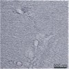 0732C 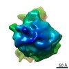 0734C C: 同じ文献を引用 ( |
|---|---|
| 電子顕微鏡画像生データ | EMPIAR-10302 (タイトル: Cryo electron tomography of spinach leaf tissue Data size: 2.2 Data #1: Unaligned tilt series of cryo lamella of spinach leaf [tilt series]) |
-リンク
| EMDBのページ | EMDB (EBI/PDBe) / EMDataResource |
|---|
-マップ
| ファイル | ダウンロード / ファイル: emd_0733.map.gz / 形式: CCP4 / 大きさ: 883.6 MB / タイプ: IMAGE STORED AS FLOATING POINT NUMBER (4 BYTES) | ||||||||||||||||||||||||||||||||||||||||||||||||||||||||||||
|---|---|---|---|---|---|---|---|---|---|---|---|---|---|---|---|---|---|---|---|---|---|---|---|---|---|---|---|---|---|---|---|---|---|---|---|---|---|---|---|---|---|---|---|---|---|---|---|---|---|---|---|---|---|---|---|---|---|---|---|---|---|
| 注釈 | The pixel size of original image is 2.65, this reconstructed tomogram has a binning factor of 4. | ||||||||||||||||||||||||||||||||||||||||||||||||||||||||||||
| 投影像・断面図 | 画像のコントロール
画像は Spider により作成 これらの図は立方格子座標系で作成されたものです | ||||||||||||||||||||||||||||||||||||||||||||||||||||||||||||
| ボクセルのサイズ | X=Y=Z: 10.6 Å | ||||||||||||||||||||||||||||||||||||||||||||||||||||||||||||
| 密度 |
| ||||||||||||||||||||||||||||||||||||||||||||||||||||||||||||
| 対称性 | 空間群: 1 | ||||||||||||||||||||||||||||||||||||||||||||||||||||||||||||
| 詳細 | EMDB XML:
CCP4マップ ヘッダ情報:
| ||||||||||||||||||||||||||||||||||||||||||||||||||||||||||||
 Z (Sec.)
 Y (Row.)
 X (Col.)


-添付データ
- 試料の構成要素
-全体 : cryo-lamella of spinach leaf
| 全体 | 名称: cryo-lamella of spinach leaf |
|---|---|
| 要素 |
|
-超分子 #1: cryo-lamella of spinach leaf
| 超分子 | 名称: cryo-lamella of spinach leaf / タイプ: tissue / ID: 1 / 親要素: 0 詳細: A puncher was used to cut a circular slice of leaf at about 2 mm diameter. The slice was then fitting into carrier for high pressure freezing. |
|---|---|
| 由来(天然) | 生物種: Spinacia oleracea (ホウレンソウ) |
-実験情報
-構造解析
| 手法 | クライオ電子顕微鏡法 |
|---|---|
 解析 解析 | 電子線トモグラフィー法 |
| 試料の集合状態 | tissue |
-試料調製
| 緩衝液 | pH: 7 / 詳細: phosphate buffered saline (PBS) |
|---|---|
| 凍結 | 凍結剤: NITROGEN |
| 詳細 | Leaf slice was put in the recess of the carrier and cryoprotectant 1-hexadecene was added to fill the surrounding area. Then a sapphire disk was loaded on top of the carrier before the whole composed sandwich was frozen. |
| 加圧凍結法 | 装置: OTHER 詳細: Leaf slice was put in the recess of the carrier and cryoprotectant 1-hexadecene was added to fill the surrounding area. Then a sapphire disk was loaded on top of the carrier before the whole ...詳細: Leaf slice was put in the recess of the carrier and cryoprotectant 1-hexadecene was added to fill the surrounding area. Then a sapphire disk was loaded on top of the carrier before the whole composed sandwich was frozen.. The value given for _emd_high_pressure_freezing.instrument is HPF COMPACT 01. This is not in a list of allowed values {'BAL-TEC HPM 010', 'LEICA EM HPM100', 'OTHER', 'LEICA EM PACT2', 'LEICA EM PACT', 'EMS-002 RAPID IMMERSION FREEZER'} so OTHER is written into the XML file. |
| Cryo protectant | 1-hexadecene |
| 切片作成 | 集束イオンビーム - 装置: OTHER / 集束イオンビーム - イオン: OTHER / 集束イオンビーム - 電圧: 30 kV / 集束イオンビーム - 電流: 0.08 nA / 集束イオンビーム - 時間: 3600 sec. / 集束イオンビーム - 温度: 93 K / 集束イオンビーム - Initial thickness: 200 nm / 集束イオンビーム - 最終 厚さ: 17 nm 集束イオンビーム - 詳細: Then the carrier was transfer with the cryo-transfer shuttle into the SEM chamber by using Quorum PP3000T cryotransfer system under -180 degree. To improve sample ...集束イオンビーム - 詳細: Then the carrier was transfer with the cryo-transfer shuttle into the SEM chamber by using Quorum PP3000T cryotransfer system under -180 degree. To improve sample conductivity and reduce curtaining artifacts, the samples were deposited with organometallic platinum using the in situ gas injection system (GIS) operated at 5 seconds gas injection time before milling. During the cryo-FIB milling process, the milling angle is nearly in parallel with the carrier, and the milling was performed parallel from both sides of the sample platform to produce lamella. Rough milling is produced with the accelerating voltage of the ion beam at 30 kV, and current at 0.79 nA-0.43 nA. The initial milling width is about 20 um and depth is about 20 um. To facilitate tomography data collection, ice at the notch above lamella was removed to get a trapezoid-shaped milling pattern. After rough milling, one side of the lamella is jagged from the main platform. When the thickness of lamella reaches about 1 um the ion current is reduced to 0.23 nA or 80 pA until thickness finally reaching 150 to 250 nm.. The value given for _emd_sectioning_focused_ion_beam.instrument is Helios NanoLab 600i. This is not in a list of allowed values {'DB235', 'OTHER'} so OTHER is written into the XML file. |
- 電子顕微鏡法
| 顕微鏡 | FEI TITAN KRIOS |
|---|---|
| 撮影 | フィルム・検出器のモデル: GATAN K2 SUMMIT (4k x 4k) 検出モード: COUNTING / デジタル化 - サイズ - 横: 3838 pixel / デジタル化 - サイズ - 縦: 3710 pixel / デジタル化 - 画像ごとのフレーム数: 1-21 / 平均露光時間: 1.0 sec. / 平均電子線量: 3.0 e/Å2 |
| 電子線 | 加速電圧: 300 kV / 電子線源:  FIELD EMISSION GUN FIELD EMISSION GUN |
| 電子光学系 | 照射モード: FLOOD BEAM / 撮影モード: BRIGHT FIELD / Cs: 2.7 mm / 最大 デフォーカス(公称値): 6.0 µm / 最小 デフォーカス(公称値): 5.0 µm |
| 試料ステージ | 試料ホルダーモデル: FEI TITAN KRIOS AUTOGRID HOLDER ホルダー冷却材: NITROGEN |
| 実験機器 |  モデル: Titan Krios / 画像提供: FEI Company |
-画像解析
| 最終 再構成 | アルゴリズム: BACK PROJECTION / ソフトウェア - 名称: IMOD (ver. 4.9.2) / 使用した粒子像数: 42 |
|---|
