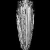
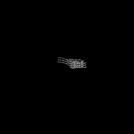
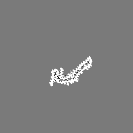
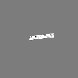
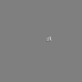
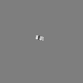
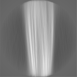
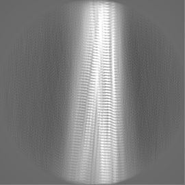
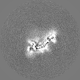
- EMDB-0077: Narrow Pick Filament from Pick's disease brain -
+
Open data
ID or keywords:
Loading...
-
Basic information
Entry
Database: EMDB / ID: EMD-0077
Title
Narrow Pick Filament from Pick's disease brain
Map data
Sample
Tissue: Tau filaments extracted from the frontotemporal cortex of a patient with Pick's disease
Protein or peptide: Microtubule-associated protein tau
Keywords
tau protein / tau / Pick's disease / tauopathy / neurodegenerative disease / proteinopathy / amyloid / filament / helical / Pick body / fibril / neurodegeneration / MAPT / microtubule-associated protein tau / PROTEIN FIBRIL
Function / homology
Function and homology information
plus-end-directed organelle transport along microtubule / histone-dependent DNA binding / negative regulation of protein localization to mitochondrion / neurofibrillary tangle / microtubule lateral binding / axonal transport / tubulin complex / positive regulation of protein localization to synapse / phosphatidylinositol bisphosphate binding / generation of neurons ...plus-end-directed organelle transport along microtubule / histone-dependent DNA binding / negative regulation of protein localization to mitochondrion / neurofibrillary tangle / microtubule lateral binding / axonal transport / tubulin complex / positive regulation of protein localization to synapse / phosphatidylinositol bisphosphate binding / generation of neurons / rRNA metabolic process / axonal transport of mitochondrion / regulation of mitochondrial fission / axon development / regulation of microtubule-based movement / intracellular distribution of mitochondria / regulation of chromosome organization / central nervous system neuron development / minor groove of adenine-thymine-rich DNA binding / lipoprotein particle binding / microtubule polymerization / negative regulation of mitochondrial membrane potential / regulation of microtubule polymerization / dynactin binding / apolipoprotein binding / protein polymerization / main axon / Caspase-mediated cleavage of cytoskeletal proteins / regulation of microtubule polymerization or depolymerization / negative regulation of mitochondrial fission / axolemma / glial cell projection / neurofibrillary tangle assembly / positive regulation of axon extension / regulation of cellular response to heat / positive regulation of microtubule polymerization / positive regulation of protein localization / Activation of AMPK downstream of NMDARs / positive regulation of superoxide anion generation / cellular response to brain-derived neurotrophic factor stimulus / regulation of long-term synaptic depression / supramolecular fiber organization / cytoplasmic microtubule organization / regulation of calcium-mediated signaling / axon cytoplasm / somatodendritic compartment / synapse assembly / phosphatidylinositol binding / nuclear periphery / astrocyte activation / enzyme inhibitor activity / protein phosphatase 2A binding / stress granule assembly / regulation of microtubule cytoskeleton organization / regulation of autophagy / cellular response to reactive oxygen species / microglial cell activation / cellular response to nerve growth factor stimulus / Hsp90 protein binding / protein homooligomerization / SH3 domain binding / PKR-mediated signaling / synapse organization / regulation of synaptic plasticity / response to lead ion / microtubule cytoskeleton organization / memory / neuron projection development / cytoplasmic ribonucleoprotein granule / cell-cell signaling / single-stranded DNA binding / cellular response to heat / growth cone / protein-folding chaperone binding / microtubule cytoskeleton / actin binding / cell body / double-stranded DNA binding / sequence-specific DNA binding / microtubule binding / amyloid fibril formation / dendritic spine / microtubule / protein-macromolecule adaptor activity / learning or memory / neuron projection / membrane raft / negative regulation of gene expression / axon / neuronal cell body / DNA damage response / dendrite / protein kinase binding / enzyme binding / mitochondrion / DNA binding / RNA binding / extracellular region / identical protein binding / nucleus Similarity search - Function
Microtubule-associated protein Tau / Microtubule associated protein, tubulin-binding repeat / Tau and MAP protein, tubulin-binding repeat / Tau and MAP proteins tubulin-binding repeat signature. / Tau and MAP proteins tubulin-binding repeat profile. / : Similarity search - Domain/homology
National Institutes of Health/National Institute on Aging (NIH/NIA)
P30-AG010133
United States
European Union
Joint Programme-Neurodegeneration Research
United Kingdom
European Union
Horizon 2020 IMPRiND
United Kingdom
Citation
Journal: Nature / Year: 2018 Title: Structures of filaments from Pick's disease reveal a novel tau protein fold. Authors: Benjamin Falcon / Wenjuan Zhang / Alexey G Murzin / Garib Murshudov / Holly J Garringer / Ruben Vidal / R Anthony Crowther / Bernardino Ghetti / Sjors H W Scheres / Michel Goedert / Abstract: The ordered assembly of tau protein into abnormal filamentous inclusions underlies many human neurodegenerative diseases. Tau assemblies seem to spread through specific neural networks in each ...The ordered assembly of tau protein into abnormal filamentous inclusions underlies many human neurodegenerative diseases. Tau assemblies seem to spread through specific neural networks in each disease, with short filaments having the greatest seeding activity. The abundance of tau inclusions strongly correlates with disease symptoms. Six tau isoforms are expressed in the normal adult human brain-three isoforms with four microtubule-binding repeats each (4R tau) and three isoforms that lack the second repeat (3R tau). In various diseases, tau filaments can be composed of either 3R or 4R tau, or of both. Tau filaments have distinct cellular and neuroanatomical distributions, with morphological and biochemical differences suggesting that they may be able to adopt disease-specific molecular conformations. Such conformers may give rise to different neuropathological phenotypes, reminiscent of prion strains. However, the underlying structures are not known. Using electron cryo-microscopy, we recently reported the structures of tau filaments from patients with Alzheimer's disease, which contain both 3R and 4R tau. Here we determine the structures of tau filaments from patients with Pick's disease, a neurodegenerative disorder characterized by frontotemporal dementia. The filaments consist of residues Lys254-Phe378 of 3R tau, which are folded differently from the tau filaments in Alzheimer's disease, establishing the existence of conformers of assembled tau. The observed tau fold in the filaments of patients with Pick's disease explains the selective incorporation of 3R tau in Pick bodies, and the differences in phosphorylation relative to the tau filaments of Alzheimer's disease. Our findings show how tau can adopt distinct folds in the human brain in different diseases, an essential step for understanding the formation and propagation of molecular conformers.
Entire : Tau filaments extracted from the frontotemporal cortex of a patie...
Entire
Name: Tau filaments extracted from the frontotemporal cortex of a patient with Pick's disease
Components
Tissue: Tau filaments extracted from the frontotemporal cortex of a patient with Pick's disease
Protein or peptide: Microtubule-associated protein tau
-
Supramolecule #1: Tau filaments extracted from the frontotemporal cortex of a patie...
Supramolecule
Name: Tau filaments extracted from the frontotemporal cortex of a patient with Pick's disease type: tissue / ID: 1 / Parent: 0 / Macromolecule list: all
Fourier-space refinement of the complete atomic model against the narrow Pick filament map was performed in REFMAC. A stack of three consecutive monomers was refined to preserve nearest-neighbour interactions for the middle chain.
Refinement
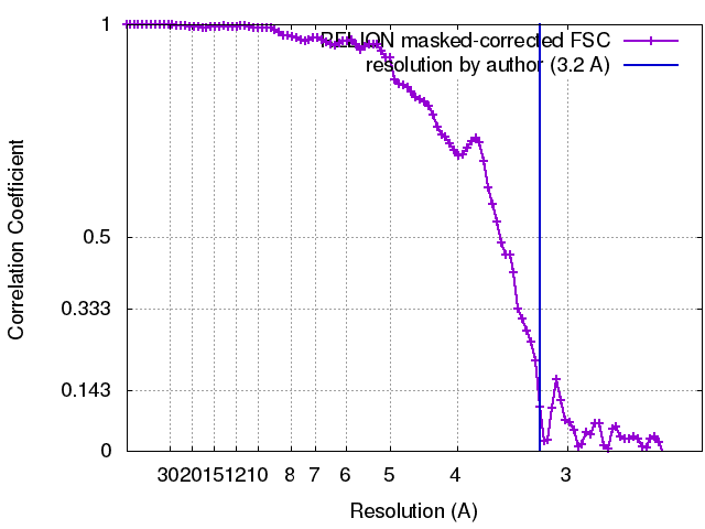
Space: RECIPROCAL / Protocol: AB INITIO MODEL / Overall B value: 57 / Target criteria: Fourier shell correlation
Output model
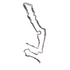
PDB-6gx5: Narrow Pick Filament from Pick's disease brain
+
About Yorodumi
-
News
-
Feb 9, 2022. New format data for meta-information of EMDB entries
New format data for meta-information of EMDB entries
Version 3 of the EMDB header file is now the official format.
The previous official version 1.9 will be removed from the archive.
In the structure databanks used in Yorodumi, some data are registered as the other names, "COVID-19 virus" and "2019-nCoV". Here are the details of the virus and the list of structure data.
Jan 31, 2019. EMDB accession codes are about to change! (news from PDBe EMDB page)
EMDB accession codes are about to change! (news from PDBe EMDB page)
The allocation of 4 digits for EMDB accession codes will soon come to an end. Whilst these codes will remain in use, new EMDB accession codes will include an additional digit and will expand incrementally as the available range of codes is exhausted. The current 4-digit format prefixed with “EMD-” (i.e. EMD-XXXX) will advance to a 5-digit format (i.e. EMD-XXXXX), and so on. It is currently estimated that the 4-digit codes will be depleted around Spring 2019, at which point the 5-digit format will come into force.
The EM Navigator/Yorodumi systems omit the EMD- prefix.
Related info.:Q: What is EMD? / ID/Accession-code notation in Yorodumi/EM Navigator
Yorodumi is a browser for structure data from EMDB, PDB, SASBDB, etc.
This page is also the successor to EM Navigator detail page, and also detail information page/front-end page for Omokage search.
The word "yorodu" (or yorozu) is an old Japanese word meaning "ten thousand". "mi" (miru) is to see.
Related info.:EMDB / PDB / SASBDB / Comparison of 3 databanks / Yorodumi Search / Aug 31, 2016. New EM Navigator & Yorodumi / Yorodumi Papers / Jmol/JSmol / Function and homology information / Changes in new EM Navigator and Yorodumi
 Movie
Movie Controller


 Open data
 Basic information
 Map data
 Sample
 Keywords
 Function and homology information
 Homo sapiens (human)
 Authors
 United Kingdom,
  United States, 6 items
  Citation
 Structure visualization
 Downloads & links
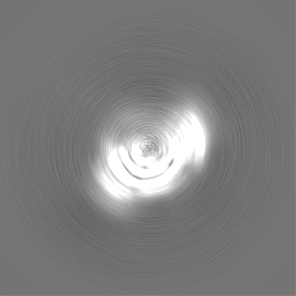
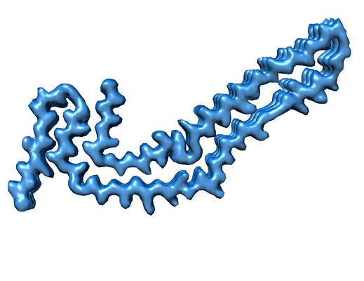 emd_0077.png
 http://ftp.pdbj.org/pub/emdb/structures/EMD-0077


 Z (Sec.)
 Y (Row.)
 X (Col.)


 Sample components
 Processing
 Electron microscopy
 FIELD EMISSION GUN