 Movie
Movie Controller


+ Open data

- Basic information
| Entry | 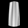 | ||||||||||||||||||
|---|---|---|---|---|---|---|---|---|---|---|---|---|---|---|---|---|---|---|---|
| Title | Four protofilament beta-2-microglobulin amyloid fibril | ||||||||||||||||||
 Map data Map data | Four protofilament beta-2-microglobulin amyloid fibril | ||||||||||||||||||
 Sample Sample |
| ||||||||||||||||||
| Biological species |  Homo sapiens (human) Homo sapiens (human) | ||||||||||||||||||
| Method | helical reconstruction / cryo EM / Resolution: 9.42 Å | ||||||||||||||||||
 Authors Authors | Iadanza MG / Ranson NA | ||||||||||||||||||
| Funding support |  United Kingdom, United Kingdom,  United States, 5 items United States, 5 items
| ||||||||||||||||||
 Citation Citation | Journal: Nat Commun / Year: 2018 Title: The structure of a β-microglobulin fibril suggests a molecular basis for its amyloid polymorphism. Authors: Matthew G Iadanza / Robert Silvers / Joshua Boardman / Hugh I Smith / Theodoros K Karamanos / Galia T Debelouchina / Yongchao Su / Robert G Griffin / Neil A Ranson / Sheena E Radford / Abstract: All amyloid fibrils contain a cross-β fold. How this structure differs in fibrils formed from proteins associated with different diseases remains unclear. Here, we combine cryo-EM and MAS-NMR to ...All amyloid fibrils contain a cross-β fold. How this structure differs in fibrils formed from proteins associated with different diseases remains unclear. Here, we combine cryo-EM and MAS-NMR to determine the structure of an amyloid fibril formed in vitro from β-microglobulin (βm), the culprit protein of dialysis-related amyloidosis. The fibril is composed of two identical protofilaments assembled from subunits that do not share βm's native tertiary fold, but are formed from similar β-strands. The fibrils share motifs with other amyloid fibrils, but also contain unique features including π-stacking interactions perpendicular to the fibril axis and an intramolecular disulfide that stabilises the subunit fold. We also describe a structural model for a second fibril morphology and show that it is built from the same subunit fold. The results provide insights into the mechanisms of fibril formation and the commonalities and differences within the amyloid fold in different protein sequences. | ||||||||||||||||||
| History |
|
- Structure visualization
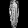
| Structure viewer | EM map:  SurfViewMolmilJmol/JSmol SurfViewMolmilJmol/JSmol |
|---|---|
| Supplemental images |

- Downloads & links
-EMDB archive
| Map data | emd_0019.map.gz | 303.9 MB | EMDB map data format | |
|---|---|---|---|---|
| Header (meta data) | emd-0019-v30.xmlemd-0019.xml | 14.4 KB 14.4 KB | Display Display | EMDB header |
| Images | 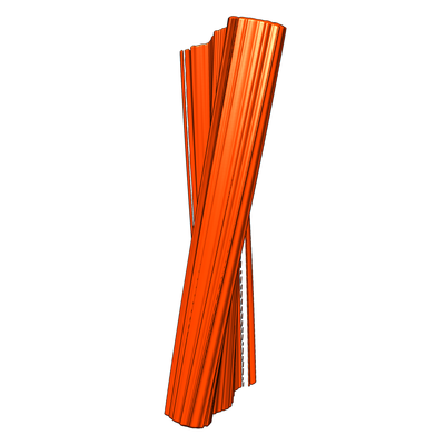 emd_0019.png emd_0019.png | 45.6 KB | ||
| Archive directory |  http://ftp.pdbj.org/pub/emdb/structures/EMD-0019ftp://ftp.pdbj.org/pub/emdb/structures/EMD-0019 http://ftp.pdbj.org/pub/emdb/structures/EMD-0019ftp://ftp.pdbj.org/pub/emdb/structures/EMD-0019 | HTTPS FTP |
-Related structure data
| Similar structure data |
|---|
-Links
| EMDB pages | EMDB (EBI/PDBe) / EMDataResource |
|---|---|
| Related items in Molecule of the Month |

-Map
| File | Download / File: emd_0019.map.gz / Format: CCP4 / Size: 824 MB / Type: IMAGE STORED AS FLOATING POINT NUMBER (4 BYTES) | ||||||||||||||||||||||||||||||||||||
|---|---|---|---|---|---|---|---|---|---|---|---|---|---|---|---|---|---|---|---|---|---|---|---|---|---|---|---|---|---|---|---|---|---|---|---|---|---|
| Annotation | Four protofilament beta-2-microglobulin amyloid fibril | ||||||||||||||||||||||||||||||||||||
| Projections & slices | Image control
Images are generated by Spider. | ||||||||||||||||||||||||||||||||||||
| Voxel size | X=Y=Z: 1.06 Å | ||||||||||||||||||||||||||||||||||||
| Density |
| ||||||||||||||||||||||||||||||||||||
| Symmetry | Space group: 1 | ||||||||||||||||||||||||||||||||||||
| Details | EMDB XML:
|
 Z (Sec.)
 Y (Row.)
 X (Col.)


-Supplemental data
- Sample components
-Entire : four protofilament beta-2-microglobulin amyloid fibril
| Entire | Name: four protofilament beta-2-microglobulin amyloid fibril |
|---|---|
| Components |
|
-Supramolecule #1: four protofilament beta-2-microglobulin amyloid fibril
| Supramolecule | Name: four protofilament beta-2-microglobulin amyloid fibril type: complex / ID: 1 / Parent: 0 / Macromolecule list: all |
|---|---|
| Source (natural) | Organism: Homo sapiens (human) |
| Recombinant expression | Organism:  |
| Molecular weight | Theoretical: 11.7 KDa |
-Macromolecule #1: Human beta-2-microglobulin
| Macromolecule | Name: Human beta-2-microglobulin / type: protein_or_peptide / ID: 1 / Enantiomer: LEVO |
|---|---|
| Source (natural) | Organism: Homo sapiens (human) |
| Recombinant expression | Organism: |
| Sequence | String: IQRTPKIQVY SRHPAENGKS NFLNCYVSGF HPSDIEVDLL KNGERIEKVE HSDLSFSKDW SFYLLYYTE FTPTEKDEYA CRVNHVTLSQ PKIVKWDRDM |
-Experimental details
-Structure determination
| Method | cryo EM |
|---|---|
 Processing Processing | helical reconstruction |
| Aggregation state | filament |
-Sample preparation
| Concentration | 0.025 mg/mL | ||||||||||||
|---|---|---|---|---|---|---|---|---|---|---|---|---|---|
| Buffer | pH: 2.5 Component:
| ||||||||||||
| Grid | Model: Quantifoil R3.5/1 / Material: COPPER / Mesh: 400 / Support film - Material: CARBON / Support film - topology: HOLEY / Pretreatment - Type: PLASMA CLEANING | ||||||||||||
| Vitrification | Cryogen name: ETHANE / Chamber humidity: 80 % / Chamber temperature: 281.15 K / Instrument: FEI VITROBOT MARK II | ||||||||||||
| Details | Quiescent growth at 0.25 mg/ml for 5 weeks, diluted 10x with buffer |
- Electron microscopy
| Microscope | FEI TITAN KRIOS |
|---|---|
| Image recording | Film or detector model: GATAN K2 SUMMIT (4k x 4k) / Detector mode: COUNTING / Digitization - Frames/image: 3-40 / Number grids imaged: 1 / Number real images: 5549 / Average exposure time: 10.0 sec. / Average electron dose: 38.5 e/Å2 |
| Electron beam | Acceleration voltage: 300 kV / Electron source:  FIELD EMISSION GUN FIELD EMISSION GUN |
| Electron optics | C2 aperture diameter: 100.0 µm / Illumination mode: FLOOD BEAM / Imaging mode: BRIGHT FIELD / Cs: 2.7 mm / Nominal defocus max: 0.00325 µm / Nominal defocus min: 0.00175 µm / Nominal magnification: 130000 |
| Sample stage | Specimen holder model: FEI TITAN KRIOS AUTOGRID HOLDER / Cooling holder cryogen: NITROGEN |
| Experimental equipment |  Model: Titan Krios / Image courtesy: FEI Company |
-Image processing
| Final reconstruction | Number classes used: 1 Applied symmetry - Helical parameters - Δz: 2.3 Å Applied symmetry - Helical parameters - Δ&Phi: -0.161 ° Applied symmetry - Helical parameters - Axial symmetry: C1 (asymmetric) Algorithm: FOURIER SPACE / Resolution.type: BY AUTHOR / Resolution: 9.42 Å / Resolution method: FSC 0.5 CUT-OFF / Software - Name: RELION / Details: Resolution estimated using rmeasure software / Number images used: 8891 |
|---|---|
| CTF correction | Software: (Name: RELION (ver. 2.1), Gctf) |
| Startup model | Type of model: OTHER Details: Double protofilament map from same study Filtered to 60 Angstrom resolution |
| Final angle assignment | Type: NOT APPLICABLE / Software - Name: RELION (ver. 2.1) |
