Glycoside hydrolase, family 14B, plant / Glycoside hydrolase, family 14, conserved site / Beta-amylase active site 1. / Glycoside hydrolase, family 14 / Glycosyl hydrolase family 14 / Glycoside hydrolase superfamily 類似検索 - ドメイン・相同性
設備名称: Advanced Light Source (ALS) 12.3.1 (SIBYLS) / 地域: Berkeley, CA / 国: USA / 線源: X-ray synchrotron / 波長: 0.127 Å / スペクトロメータ・検出器間距離: 2 mm
検出器
名称: Pilatus3 X 2M / Pixsize x: 172 mm
スキャン
タイトル: Beta-amylase 2, chloroplastic (AtBAM2) Ndel1 / 測定日: 2019年6月11日 / 保管温度: 10 °C / セル温度: 10 °C / 照射時間: 0.3 sec. / フレーム数: 50 / 単位: 1/A /
Min
Max
Q
0.0137
0.3977
距離分布関数 P(R)
ソフトウェア P(R): GNOM 5.0 / ポイント数: 303 /
Min
Max
Q
0.013658
0.181999
P(R) point
1
303
R
0
109.8
結果
カーブのタイプ: merged コメント: We expressed Arabidopsis thaliana AtBAM2 in E. coli BL21 cells from a pETDuet-1 expression vector with an N-terminal 6-His tag as constructed by (Monroe et al., 2017, 2018). We induced ...コメント: We expressed Arabidopsis thaliana AtBAM2 in E. coli BL21 cells from a pETDuet-1 expression vector with an N-terminal 6-His tag as constructed by (Monroe et al., 2017, 2018). We induced AtBAM2 expression in BL21 E coli at OD600 with 0.3 mM IPTG in 2xYT broth with 60 µg/ml ampicillin at 30 oC overnight. Cells were lysed and sonicated in a buffer containing 50 mM NaH2PO4, pH 8, 500 mM NaCl, and 2 mM imidazole. The supernatant was loaded onto a TALON cobalt column using an AKTA Start and washed with a buffer containing 50 mM HEPES pH 8, 500 mM NaCl, 5% glycerol, 10 mM TCEP, and 40 mM imidazole. The protein was then eluted with buffer containing 50 mM HEPES pH 8, 500 mM NaCl, 5% glycerol, 10 mM TCEP, and 500 mM imidazole. Pure protein, as determined by a band present at ∼50 kDa on a 4–20% Tris‐Glycine gel stained with Coomassie Blue, was concentrated in a Spin‐X UF concentrator with a 5000 MWCO. The concentrated protein was then further purified using a HiLoad 16/60 column filled with Superdex 200 in 50 mM HEPES, pH 7. Pure protein based on SDS-PAGE was concentrated as before and the concentration of protein was determined via absorbance at 280 nm using an extinction coefficient of 94310 M−1 cm−1 which was calculated from the sequence using ProtParam (Gasteiger et al., 2005). Construction of AtBAM2-Ndel2 was constructed using a similar strategy as that of AtBAM2-Ndel1 (Monroe et al., 2017) but using the primer 5’- TCGAAGAGCGTGATTTTGCGGATCCAGCGTGTGTTCCTGTATATG-3’ and the complementary sequence. AtBAM2 with the Ndel1 or Ndel2 truncations were purified using the same scheme described for the wild-type AtBAM2.
Matching buffer exposures were collected before and after samples to ensure there was no difference in the scattering due to contamination of the sample cell. Scattering data were subtracted from buffer and then processed in PRIMUS (ATSAS 2.8.4 r10553) to create an average data file (Konarev et al., 2003). Data were analyzed using PRIMUS, GNOM, and SCÅTTER (v3.0g) to determine dimensions and create a merged data file from the two data sets at 30 and 50 µM AtBAM2 (Konarev et al., 2003). We then used the merged data file in DAMMIF (v1.1.2) and DAMMIN (v5.3) to generate the dummy-atom model and aligned the result to the all-atom structure using SASTBX (Svergun, 1992, 1999; Svergun et al., 2001; Franke & Svergun, 2009; Liu et al., 2012).
 ムービー
ムービー コントローラー


 データを開く
 基本情報
基本情報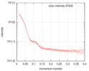
 試料
試料 機能・相同性情報
 引用
引用 登録者
登録者 構造の表示
構造の表示 ダウンロードとリンク
 SASDGZ4
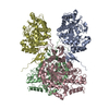
 / 線源: X-ray synchrotron / 波長: 0.127 Å / スペクトロメータ・検出器間距離: 2 mm


