[English] 日本語
 Yorodumi
- SASDFZ4: R16-24 human dystrophin fragment (Human dystrophin central domain... -
+ Open data
- Basic information
| Entry |  |
|---|---|
 Sample Sample | R16-24 human dystrophin fragment
|
| Function / homology |  Function and homology information Function and homology informationregulation of muscle system process / regulation of cellular response to growth factor stimulus / syntrophin complex / cardiac muscle cell action potential / regulation of skeletal muscle contraction / dystrophin-associated glycoprotein complex / synaptic signaling / cell-substrate junction / peptide biosynthetic process / motile cilium assembly ...regulation of muscle system process / regulation of cellular response to growth factor stimulus / syntrophin complex / cardiac muscle cell action potential / regulation of skeletal muscle contraction / dystrophin-associated glycoprotein complex / synaptic signaling / cell-substrate junction / peptide biosynthetic process / motile cilium assembly / dystroglycan binding / regulation of skeletal muscle contraction by regulation of release of sequestered calcium ion / vinculin binding / regulation of sodium ion transmembrane transport / Formation of the dystrophin-glycoprotein complex (DGC) / costamere / muscle cell development / regulation of calcium ion transmembrane transport / neuron projection terminus / Striated Muscle Contraction / filopodium membrane / structural constituent of muscle / muscle organ development / muscle cell cellular homeostasis / myosin binding / maintenance of blood-brain barrier / nitric-oxide synthase binding / Non-integrin membrane-ECM interactions / neuron development / regulation of cardiac muscle contraction by regulation of the release of sequestered calcium ion / skeletal muscle tissue development / cardiac muscle contraction / regulation of release of sequestered calcium ion into cytosol by sarcoplasmic reticulum / response to muscle stretch / positive regulation of neuron differentiation / regulation of heart rate / filopodium / positive regulation of neuron projection development / sarcolemma / structural constituent of cytoskeleton / Z disc / intracellular protein localization / actin binding / protein-containing complex assembly / postsynaptic membrane / cytoskeleton / membrane raft / synapse / cell surface / protein-containing complex / zinc ion binding / nucleus / plasma membrane / cytosol Similarity search - Function |
 Contact author Contact author |
|
- Structure visualization
- Downloads & links
-Data source
| SASBDB page |  SASDFZ4 SASDFZ4 |
|---|
-Related structure data
| Similar structure data | Similarity search - Function & homology  F&H Search F&H Search |
|---|
- External links
| Related items in Molecule of the Month |
|---|
-Models
- Sample
 Sample Sample | Name: R16-24 human dystrophin fragment |
|---|---|
| Buffer | Name: NaP 20 mM, NaCl 300 mM, EDTA 1 mM, Glycérol 2% / pH: 7.5 |
| Entity #1557 | Type: protein Description: Human dystrophin central domain R16-24 fragment Formula weight: 126.616 / Num. of mol.: 1 / References: UniProt: P11532 Sequence: MSYYHHHHHH DYDIPTTENL YFOGAMGRSL VPRGSLEISY VPSTYLTEIT HVSQALLEVE QLLNAPDLCA KDFEDLFKQE ESLKNIKDSL QQSSGRIDII HSKKTAALQS ATPVERVKLQ EALSQLDFQW EKVNKMYKDR QGRFDRSVEK WRRFHYDIKI FNQWLTEAEQ ...Sequence: MSYYHHHHHH DYDIPTTENL YFOGAMGRSL VPRGSLEISY VPSTYLTEIT HVSQALLEVE QLLNAPDLCA KDFEDLFKQE ESLKNIKDSL QQSSGRIDII HSKKTAALQS ATPVERVKLQ EALSQLDFQW EKVNKMYKDR QGRFDRSVEK WRRFHYDIKI FNQWLTEAEQ FLRKTQIPEN WEHAKYKWYL KELQDGIGQR QTVVRTLNAT GEEIIQQSSK TDASILQEKL GSLNLRWQEV CKQLSDRKKR LEEQKNILSE FQRDLNEFVL WLEEADNIAS IPLEPGKEQQ LKEKLEQVKL LVEELPLRQA GILKQLNETG GPVLVSAPIS PEEQDKLENK LKQTNLQWIK VSRALPEKQG EIEAQIKDLG QLEKKLEDLE EQLNHLLLWL SPIRNQLEIY NQPNQEGPFD VQETEIAVQA KQPDVEEILS KGQHLYKEKP ATQPVKRKLE DLSSEWKAVN RLLQELRAKQ PDLAPGLTTI GASPTQTVTL VTQPVVTKET AISKLEMPSS LMLEVPALAD FNRAWTELTD WLSLLDQVIK SQRVMVGDLE DINEMIIKQK ATMQDLEQRR PQLEELITAA QNLKNKTSNQ EARTIITDRI ERIQNQWDEV QEHLQNRRQQ LNEMLKDSTQ WLEAKEEAEQ VLGQARAKLE SWKEGPYTVD AIQKKITETK QLAKDLRQWQ TNVDVANDLA LKLLRDYSAD DTRKVHMITE NINASWRSIH KRVSEREAAL EETHRLLQQF PLDLEKFLAW LTEAETTANV LQDATRKERL LEDSKGVKEL MKQWQDLQGE IEAHTDVYHN LDENSQKILR SLEGSDDAVL LQRRLDNMNF KWSELRKKSL NIRSHLEASS DQWKRLHLSL QELLVWLQLK DDELSRQAPI GGDFPAVQKQ NDVHRAFKRE LKTKEPVIMS TLETVRIFLT EQPLEGLEKL YQEPRELPPE ERAQNVTRLL RKQAEEVNTE WEKLNLHSAD WQRKIDETLE RLQELQEATD ELDLKLRQAE VIKGSWQPVG DLLIDSLQDH LEKVKALRGE IAPLKENVSH VNDLARQLTT LGIQLSPYNL STLEDLNTRW KLLQVAVEDR VRQLHE |
-Experimental information
| Beam | Instrument name: SOLEIL SWING  / City: Saint-Aubin / 国: France / City: Saint-Aubin / 国: France  / Type of source: X-ray synchrotron / Wavelength: 0.1033 Å / Dist. spec. to detc.: 2 mm / Type of source: X-ray synchrotron / Wavelength: 0.1033 Å / Dist. spec. to detc.: 2 mm | ||||||||||||
|---|---|---|---|---|---|---|---|---|---|---|---|---|---|
| Detector | Name: AVIEX PCCD170170 / Type: CCD | ||||||||||||
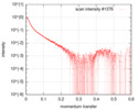
| Scan |
| ||||||||||||
| Distance distribution function P(R) |
| ||||||||||||
| Result | Comments: SEC-SAXS was performed at 20°C using the following parameters: Column: BioSEC5-500Å (4.6 mm id * 300 mm); Flow rate: 0.3 mL/min; Sample injection concentration: 4.5 mg/mL; Injection ...Comments: SEC-SAXS was performed at 20°C using the following parameters: Column: BioSEC5-500Å (4.6 mm id * 300 mm); Flow rate: 0.3 mL/min; Sample injection concentration: 4.5 mg/mL; Injection volume: 50μL. The data were collected through the SEC peak of the protein as a series of 38 x 0.75 second exposures. The experimental molecular weight was determined from SEC-MALS (130 kDa).
|
 Movie
Movie Controller


