Instrument name: SOLEIL SWING / City: Saint-Aubin / 国: France / Type of source: X-ray synchrotron / Wavelength: 0.1033 Å / Dist. spec. to detc.: 2 mm
Detector
Name: AVIEX PCCD170170 / Type: CCD
Scan
Title: R4-15 human dystrophin fragment / Measurement date: May 26, 2016 / Cell temperature: 20 °C / Exposure time: 1 sec. / Number of frames: 21 / Unit: 1/A /
Min
Max
Q
0.0051
0.5496
Distance distribution function P(R)
Sofotware P(R): GNOM 4.6 / Number of points: 444 /
Min
Max
Q
0.008115
0.2327
P(R) point
6
449
R
0
475
Result
Experimental MW: 150 kDa / D max: 47.5 / Type of curve: sec Comments: SEC-SAXS was performed at 20°C using the following parameters: Column: BioSEC5-500Å (4.6 mm id * 300 mm); Flow rate: 0.3 mL/min; Sample injection concentration: 4.4 mg/mL; Injection ...Comments: SEC-SAXS was performed at 20°C using the following parameters: Column: BioSEC5-500Å (4.6 mm id * 300 mm); Flow rate: 0.3 mL/min; Sample injection concentration: 4.4 mg/mL; Injection volume: 50μL. The data were collected through the SEC peak of the protein as a series of 21 x 1 second exposures.
The experimental molecular weight was determined from SEC-MALS (150 kDa).
P(R)
Guinier
Forward scattering, I0
1.742
1.53
Radius of gyration, Rg
12.57 nm
9.69 nm
+
About Yorodumi
-
News
-
Feb 9, 2022. New format data for meta-information of EMDB entries
New format data for meta-information of EMDB entries
Version 3 of the EMDB header file is now the official format.
The previous official version 1.9 will be removed from the archive.
In the structure databanks used in Yorodumi, some data are registered as the other names, "COVID-19 virus" and "2019-nCoV". Here are the details of the virus and the list of structure data.
Jan 31, 2019. EMDB accession codes are about to change! (news from PDBe EMDB page)
EMDB accession codes are about to change! (news from PDBe EMDB page)
The allocation of 4 digits for EMDB accession codes will soon come to an end. Whilst these codes will remain in use, new EMDB accession codes will include an additional digit and will expand incrementally as the available range of codes is exhausted. The current 4-digit format prefixed with “EMD-” (i.e. EMD-XXXX) will advance to a 5-digit format (i.e. EMD-XXXXX), and so on. It is currently estimated that the 4-digit codes will be depleted around Spring 2019, at which point the 5-digit format will come into force.
The EM Navigator/Yorodumi systems omit the EMD- prefix.
Related info.:Q: What is EMD? / ID/Accession-code notation in Yorodumi/EM Navigator
Yorodumi is a browser for structure data from EMDB, PDB, SASBDB, etc.
This page is also the successor to EM Navigator detail page, and also detail information page/front-end page for Omokage search.
The word "yorodu" (or yorozu) is an old Japanese word meaning "ten thousand". "mi" (miru) is to see.
Related info.:EMDB / PDB / SASBDB / Comparison of 3 databanks / Yorodumi Search / Aug 31, 2016. New EM Navigator & Yorodumi / Yorodumi Papers / Jmol/JSmol / Function and homology information / Changes in new EM Navigator and Yorodumi
 Movie
Movie Controller

 Yorodumi
 Open data
 Basic information
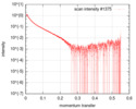
 Sample
 Function and homology information
 Contact author
 Structure visualization
 Downloads & links
 SASDFY4


 / City: Saint-Aubin / 国: France


