[English] 日本語
Yorodumi
- SASDFT4: Conformation of the R11-15 human dystrophin fragment (SANS) -

+
Open data


ID or keywords:

Loading...

-
Basic information

Entry
Database: SASBDB / ID: SASDFT4
SampleConformation of the R11-15 human dystrophin fragment (SANS)
  • Dystrophin (R11-15 human dystrophin fragment) (protein), Dystrophin 1461-1973, Homo sapiens
Function / homology
Function and homology information


regulation of muscle system process / regulation of cellular response to growth factor stimulus / regulation of skeletal muscle contraction / syntrophin complex / synaptic signaling / cardiac muscle cell action potential / regulation of voltage-gated calcium channel activity / negative regulation of peptidyl-cysteine S-nitrosylation / positive regulation of sodium ion transmembrane transporter activity / dystrophin-associated glycoprotein complex ...regulation of muscle system process / regulation of cellular response to growth factor stimulus / regulation of skeletal muscle contraction / syntrophin complex / synaptic signaling / cardiac muscle cell action potential / regulation of voltage-gated calcium channel activity / negative regulation of peptidyl-cysteine S-nitrosylation / positive regulation of sodium ion transmembrane transporter activity / dystrophin-associated glycoprotein complex / regulation of skeletal muscle contraction by regulation of release of sequestered calcium ion / peptide biosynthetic process / cell-substrate junction / motile cilium assembly / dystroglycan binding / vinculin binding / muscle cell development / costamere / neuron projection terminus / Striated Muscle Contraction / filopodium membrane / muscle organ development / structural constituent of muscle / muscle cell cellular homeostasis / maintenance of blood-brain barrier / myosin binding / nitric-oxide synthase binding / Non-integrin membrane-ECM interactions / neuron development / regulation of cardiac muscle contraction by regulation of the release of sequestered calcium ion / negative regulation of peptidyl-serine phosphorylation / regulation of release of sequestered calcium ion into cytosol by sarcoplasmic reticulum / skeletal muscle tissue development / regulation of ryanodine-sensitive calcium-release channel activity / cardiac muscle contraction / response to muscle stretch / positive regulation of neuron differentiation / regulation of heart rate / filopodium / sarcolemma / protein localization / structural constituent of cytoskeleton / Z disc / positive regulation of neuron projection development / actin binding / postsynaptic membrane / protein-containing complex assembly / cytoskeleton / membrane raft / synapse / cell surface / protein-containing complex / zinc ion binding / nucleus / plasma membrane / cytosol
Similarity search - Function
Dystrophin/utrophin / EF-hand domain, type 1 / EF-hand domain, type 2 / EF hand / EF-hand / Spectrin repeat / Spectrin repeat / Spectrin/alpha-actinin / Actinin-type actin-binding domain signature 1. / Actinin-type actin-binding domain signature 2. ...Dystrophin/utrophin / EF-hand domain, type 1 / EF-hand domain, type 2 / EF hand / EF-hand / Spectrin repeat / Spectrin repeat / Spectrin/alpha-actinin / Actinin-type actin-binding domain signature 1. / Actinin-type actin-binding domain signature 2. / Spectrin repeats / Actinin-type actin-binding domain, conserved site / Calponin homology domain / Zinc finger ZZ-type signature. / Zinc-binding domain, present in Dystrophin, CREB-binding protein. / Calponin homology (CH) domain / Zinc finger, ZZ type / Zinc finger, ZZ-type / Zinc finger, ZZ-type superfamily / Zinc finger ZZ-type profile. / Calponin homology domain / CH domain superfamily / Calponin homology (CH) domain profile. / WW domain / WW/rsp5/WWP domain signature. / WW domain superfamily / WW/rsp5/WWP domain profile. / Domain with 2 conserved Trp (W) residues / WW domain / EF-hand domain pair
Similarity search - Domain/homology
Biological speciesHomo sapiens (human)
Contact author
  • Raphael Dos Santos Morais (University of Lorraine)

-
Structure visualization

Structure viewerMolecule:
MolmilJmol/JSmol

Downloads & links

-
Models

Model #2884
Type: atomic / Chi-square value: 2.185
Search similar-shape structures of this assembly by Omokage search (details)
Model #2887
Type: dummy / Radius of dummy atoms: 1.80 A / Chi-square value: 0.643 / P-value: 0.197761
Search similar-shape structures of this assembly by Omokage search (details)

-
Sample

SampleName: Conformation of the R11-15 human dystrophin fragment (SANS)
Specimen concentration: 5.6 mg/ml
BufferName: 20 mM Tris-d11, 150 mM NaCl, 0.1 mM EDTA-d16, in 100% v/v D2O
pH: 7.1 / Comment: pD 7.5 (pH 7.1)
Entity #1527Name: Dystrophin 1461-1973 / Type: protein / Description: Dystrophin (R11-15 human dystrophin fragment) / Formula weight: 60.065 / Num. of mol.: 1 / Source: Homo sapiens / References: UniProt: P11532
Sequence: GSFQKPANFE QRLQESKMIL DEVKMHLPAL ETKSVEQEVV QSQLNHCVNL YKSLSEVKSE VEMVIKTGRQ IVQKKQTENP KELDERVTAL KLHYNELGAK VTERKQQLEK CLKLSRKMRK EMNVLTEWLA ATDMELTKRS AVEGMPSNLD SEVAWGKATQ KEIEKQKVHL ...Sequence:
GSFQKPANFE QRLQESKMIL DEVKMHLPAL ETKSVEQEVV QSQLNHCVNL YKSLSEVKSE VEMVIKTGRQ IVQKKQTENP KELDERVTAL KLHYNELGAK VTERKQQLEK CLKLSRKMRK EMNVLTEWLA ATDMELTKRS AVEGMPSNLD SEVAWGKATQ KEIEKQKVHL KSITEVGEAL KTVLGKKETL VEDKLSLLNS NWIAVTSRAE EWLNLLLEYQ KHMETFDQNV DHITKWIIQA DTLLDESEKK KPQQKEDVLK RLKAELNDIR PKVDSTRDQA ANLMANHGDH CRKLVEPQIS ELNHRFAAIS HRIKTGKASI PLKELEQFNS DIQKLLEPLE AEIQQGVNLK EEDFNKDMNE DNEGTVKELL QRGDNLQQRI TDERKREEIK IKQQLLQTKH NALKDLRSQR RKKALEISHQ WYQYKRQADD LLKCLDDIEK KLASLPEPRD ERKIKEIDRE LQKKKEELNA VRRQAEGLSE DGAAMAVEPT QIQLSKRWRE IESKFAQFRR LNFAQ

-
Experimental information

BeamInstrument name: ILL D22 / City: Grenoble / : France / Type of source: neutron source / Wavelength: 0.6 Å / Dist. spec. to detc.: 8 mm
DetectorName: 128 linear sensitive Reuter-Stokes detector / Type: 3He multidetector / Pixsize x: 0.8 mm
Scan
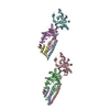
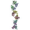
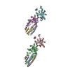
Title: Conformation of the R11-15 human dystrophin fragment (SANS)
Measurement date: Nov 1, 2016 / Cell temperature: 22 °C / Exposure time: 1200 sec. / Number of frames: 1 / Unit: 1/A /
MinMax
Q0.0065 0.6143
Distance distribution function P(R)
Sofotware P(R): GNOM 4.6 / Number of points: 118 /
MinMax
Q0.008735 0.2506
P(R) point2 119
R0 274
Result
Type of curve: single_conc
Comments: Data were collected at two sample detector positions: 1) 1.4 m for 5 min using a neutron wavelength of 0.6 nm and; 2) 8 m for 20 min using a neutron wavelength of 0.6 nm. Both datasets were ...Comments: Data were collected at two sample detector positions: 1) 1.4 m for 5 min using a neutron wavelength of 0.6 nm and; 2) 8 m for 20 min using a neutron wavelength of 0.6 nm. Both datasets were recorded on the same sample at 5.6 mg/mL, measured at 22 °C.
ExperimentalStandardPorod
MW55 kDa55 kDa-
Volume--146 nm3

P(R)GuinierGuinier error
Forward scattering, I00.2726 0.269 0.004
Radius of gyration, Rg6.47 nm6.2 nm0.2

MinMax
D-27.4
Guinier point2 10

+
About Yorodumi

-
News

-
Feb 9, 2022. New format data for meta-information of EMDB entries

New format data for meta-information of EMDB entries

  • Version 3 of the EMDB header file is now the official format.
  • The previous official version 1.9 will be removed from the archive.

Related info.:EMDB header

External links:wwPDB to switch to version 3 of the EMDB data model

-
Aug 12, 2020. Covid-19 info

Covid-19 info

URL: https://pdbj.org/emnavi/covid19.php

New page: Covid-19 featured information page in EM Navigator.

Related info.:Covid-19 info / Mar 5, 2020. Novel coronavirus structure data

+
Mar 5, 2020. Novel coronavirus structure data

Novel coronavirus structure data

Related info.:Yorodumi Speices / Aug 12, 2020. Covid-19 info

External links:COVID-19 featured content - PDBj / Molecule of the Month (242):Coronavirus Proteases

+
Jan 31, 2019. EMDB accession codes are about to change! (news from PDBe EMDB page)

EMDB accession codes are about to change! (news from PDBe EMDB page)

  • The allocation of 4 digits for EMDB accession codes will soon come to an end. Whilst these codes will remain in use, new EMDB accession codes will include an additional digit and will expand incrementally as the available range of codes is exhausted. The current 4-digit format prefixed with “EMD-” (i.e. EMD-XXXX) will advance to a 5-digit format (i.e. EMD-XXXXX), and so on. It is currently estimated that the 4-digit codes will be depleted around Spring 2019, at which point the 5-digit format will come into force.
  • The EM Navigator/Yorodumi systems omit the EMD- prefix.

Related info.:Q: What is EMD? / ID/Accession-code notation in Yorodumi/EM Navigator

External links:EMDB Accession Codes are Changing Soon! / Contact to PDBj

+
Jul 12, 2017. Major update of PDB

Major update of PDB

  • wwPDB released updated PDB data conforming to the new PDBx/mmCIF dictionary.
  • This is a major update changing the version number from 4 to 5, and with Remediation, in which all the entries are updated.
  • In this update, many items about electron microscopy experimental information are reorganized (e.g. em_software).
  • Now, EM Navigator and Yorodumi are based on the updated data.

External links:wwPDB Remediation / Enriched Model Files Conforming to OneDep Data Standards Now Available in the PDB FTP Archive

-
Yorodumi

Thousand views of thousand structures

  • Yorodumi is a browser for structure data from EMDB, PDB, SASBDB, etc.
  • This page is also the successor to EM Navigator detail page, and also detail information page/front-end page for Omokage search.
  • The word "yorodu" (or yorozu) is an old Japanese word meaning "ten thousand". "mi" (miru) is to see.

Related info.:EMDB / PDB / SASBDB / Comparison of 3 databanks / Yorodumi Search / Aug 31, 2016. New EM Navigator & Yorodumi / Yorodumi Papers / Jmol/JSmol / Function and homology information / Changes in new EM Navigator and Yorodumi

Read more