 Movie
Movie Controller


+ Open data

- Basic information
| Entry | Database: PDB / ID: 9zzm | |||||||||||||||
|---|---|---|---|---|---|---|---|---|---|---|---|---|---|---|---|---|
| Title | Two Lmod2s and incoming actin at the pointed end of F-actin | |||||||||||||||
 Components Components |
| |||||||||||||||
 Keywords Keywords | STRUCTURAL PROTEIN / actin / Lmod2 / leiomodin | |||||||||||||||
| Function / homology |  Function and homology information Function and homology informationpointed-end actin filament capping / actin nucleation / myofibril assembly / positive regulation of actin filament polymerization / sarcomere organization / M band / cytoskeletal motor activator activity / myosin heavy chain binding / tropomyosin binding / actin filament bundle ...pointed-end actin filament capping / actin nucleation / myofibril assembly / positive regulation of actin filament polymerization / sarcomere organization / M band / cytoskeletal motor activator activity / myosin heavy chain binding / tropomyosin binding / actin filament bundle / troponin I binding / filamentous actin / mesenchyme migration / myofibril / skeletal muscle myofibril / striated muscle thin filament / actin filament bundle assembly / skeletal muscle thin filament assembly / actin monomer binding / skeletal muscle fiber development / stress fiber / actin filament polymerization / titin binding / muscle contraction / actin filament organization / sarcomere / filopodium / actin filament / Hydrolases; Acting on acid anhydrides; Acting on acid anhydrides to facilitate cellular and subcellular movement / calcium-dependent protein binding / lamellipodium / actin binding / cell body / cytoskeleton / protein domain specific binding / hydrolase activity / positive regulation of gene expression / calcium ion binding / magnesium ion binding / ATP binding / identical protein binding / cytoplasm Similarity search - Function | |||||||||||||||
| Biological species |  Homo sapiens (human) Homo sapiens (human)  Oryctolagus cuniculus (rabbit) Oryctolagus cuniculus (rabbit) | |||||||||||||||
| Method | ELECTRON MICROSCOPY / single particle reconstruction / cryo EM / Resolution: 3.56 Å | |||||||||||||||
 Authors Authors | Brotzman, S.B. / Palmer, N.J. / Dominguez, R. | |||||||||||||||
| Funding support |  United States, 2items United States, 2items
| |||||||||||||||
 Citation Citation | Journal: Nat Commun / Year: 2026 Title: Mechanism of actin thin filament pointed-end elongation by leiomodin. Authors: Shayna B Brotzman / Nicholas J Palmer / Malgorzata Boczkowska / Roberto Dominguez / Abstract: In non-muscle cells, actin filaments exhibit variable lengths and rapid turnover, with subunits adding primarily at the barbed end. The situation is strikingly different in striated muscle ...In non-muscle cells, actin filaments exhibit variable lengths and rapid turnover, with subunits adding primarily at the barbed end. The situation is strikingly different in striated muscle sarcomeres, where despite rapid turnover, actin thin filaments exhibit uniform length and exchange subunits primarily at the pointed end. This filament length uniformity is tightly regulated by several proteins, including the molecular ruler nebulin in skeletal muscle and the barbed- and pointed-end capping proteins CapZ and tropomodulin (Tmod) in both skeletal and cardiac muscles. Recent studies in cells and animal models have identified leiomodin-2 (Lmod2) as an additional regulator proposed to promote pointed-end elongation to maintain thin filament length. This activity would make leiomodin the only known eukaryotic factor to drive pointed-end elongation, yet its molecular mechanism remains unresolved. Here, we present a series of cryo-electron microscopy structures that support a stepwise elongation mechanism in which two Lmod2 molecules alternate at the pointed end while recruiting actin monomers. These findings establish the molecular basis of pointed-end elongation in muscle sarcomeres and provide a framework for understanding mutations in Lmod2 that cause dilated cardiomyopathy. | |||||||||||||||
| History |
|
- Structure visualization
| Structure viewer | Molecule: MolmilJmol/JSmol |
|---|
- Downloads & links
-Download
| PDBx/mmCIF format | 9zzm.cif.gz | 459 KB | Display | PDBx/mmCIF format |
|---|---|---|---|---|
| PDB format | pdb9zzm.ent.gz | 371.9 KB | Display | PDB format |
| PDBx/mmJSON format | 9zzm.json.gz | Tree view | PDBx/mmJSON format | |
| Others |  Other downloads Other downloads |
-Validation report
| Arichive directory | https://data.pdbj.org/pub/pdb/validation_reports/zz/9zzmftp://data.pdbj.org/pub/pdb/validation_reports/zz/9zzm | HTTPS FTP |
|---|
-Related structure data
| Related structure data |  75010MC  9zziC  9zzjC C: citing same article ( M: map data used to model this data |
|---|---|
| Similar structure data |


-Links
 PDBj
PDBj


- Assembly
| Deposited unit | 
|
|---|---|
| 1 |
|
-Components
| #1: Protein | Mass: 41875.633 Da / Num. of mol.: 6 / Source method: isolated from a natural source / Source: (natural) Oryctolagus cuniculus (rabbit) / Tissue: skeletal muscleReferences: UniProt: P68135, Hydrolases; Acting on acid anhydrides; Acting on acid anhydrides to facilitate cellular and subcellular movement #2: Protein | Mass: 62865.445 Da / Num. of mol.: 2 Source method: isolated from a genetically manipulated source Source: (gene. exp.) Homo sapiens (human) / Gene: LMOD2Production host:   Escherichia coli 'BL21-Gold(DE3)pLysS AG' (bacteria) Escherichia coli 'BL21-Gold(DE3)pLysS AG' (bacteria)References: UniProt: Q6P5Q4 #3: Chemical | ChemComp-ADP /  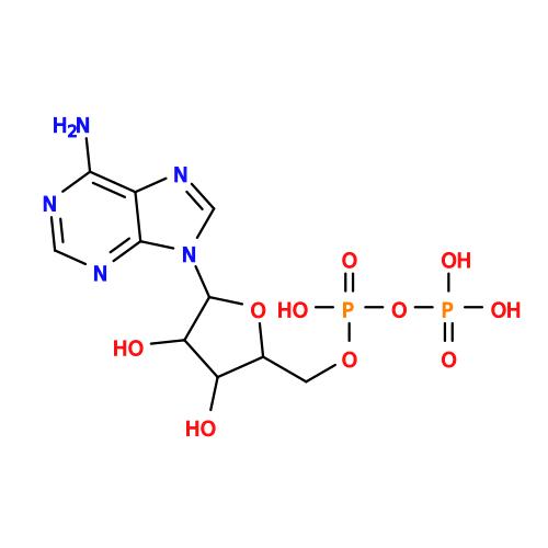 Mass: 427.201 Da / Num. of mol.: 5 / Source method: obtained synthetically / Formula: C10H15N5O10P2 / Comment: ADP, energy-carrying molecule*YM Mass: 427.201 Da / Num. of mol.: 5 / Source method: obtained synthetically / Formula: C10H15N5O10P2 / Comment: ADP, energy-carrying molecule*YM#4: Chemical | ChemComp-MG /   Mass: 24.305 Da / Num. of mol.: 6 / Source method: obtained synthetically / Formula: Mg Mass: 24.305 Da / Num. of mol.: 6 / Source method: obtained synthetically / Formula: Mg#5: Chemical | ChemComp-ATP / |  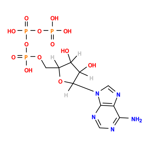 Mass: 507.181 Da / Num. of mol.: 1 / Source method: obtained synthetically / Formula: C10H16N5O13P3 / Comment: ATP, energy-carrying molecule*YM Mass: 507.181 Da / Num. of mol.: 1 / Source method: obtained synthetically / Formula: C10H16N5O13P3 / Comment: ATP, energy-carrying molecule*YMHas ligand of interest | N | Has protein modification | Y | |
|---|
-Experimental details
-Experiment
| Experiment | Method: ELECTRON MICROSCOPY |
|---|---|
| EM experiment | Aggregation state: FILAMENT / 3D reconstruction method: single particle reconstruction |
- Sample preparation
| Component |
| |||||||||||||||||||||||||||||||||||
|---|---|---|---|---|---|---|---|---|---|---|---|---|---|---|---|---|---|---|---|---|---|---|---|---|---|---|---|---|---|---|---|---|---|---|---|---|
| Molecular weight |
| |||||||||||||||||||||||||||||||||||
| Source (natural) |
| |||||||||||||||||||||||||||||||||||
| Source (recombinant) |
| |||||||||||||||||||||||||||||||||||
| Buffer solution | pH: 7.5 | |||||||||||||||||||||||||||||||||||
| Buffer component |
| |||||||||||||||||||||||||||||||||||
| Specimen | Embedding applied: NO / Shadowing applied: NO / Staining applied: NO / Vitrification applied: YES | |||||||||||||||||||||||||||||||||||
| Vitrification | Instrument: FEI VITROBOT MARK IV / Cryogen name: ETHANE / Humidity: 100 % / Chamber temperature: 277 K |
- Electron microscopy imaging
| Experimental equipment |  Model: Titan Krios / Image courtesy: FEI Company |
|---|---|
| Microscopy | Model: TFS KRIOS |
| Electron gun | Electron source:  FIELD EMISSION GUN / Accelerating voltage: 300 kV / Illumination mode: FLOOD BEAM FIELD EMISSION GUN / Accelerating voltage: 300 kV / Illumination mode: FLOOD BEAM |
| Electron lens | Mode: BRIGHT FIELD / Nominal magnification: 81000 X / Nominal defocus max: 2500 nm / Nominal defocus min: 500 nm |
| Image recording | Electron dose: 49 e/Å2 / Film or detector model: GATAN K3 (6k x 4k) |
- Processing
| EM software |
| |||||||||||||||||||||||||||||||||||
|---|---|---|---|---|---|---|---|---|---|---|---|---|---|---|---|---|---|---|---|---|---|---|---|---|---|---|---|---|---|---|---|---|---|---|---|---|
| Image processing |
| |||||||||||||||||||||||||||||||||||
| CTF correction |
| |||||||||||||||||||||||||||||||||||
| 3D reconstruction |
| |||||||||||||||||||||||||||||||||||
| Atomic model building | PDB-ID: 8F8S Accession code: 8F8S / Source name: PDB / Type: experimental model |

